Microtia
Microtia
What is microtia?
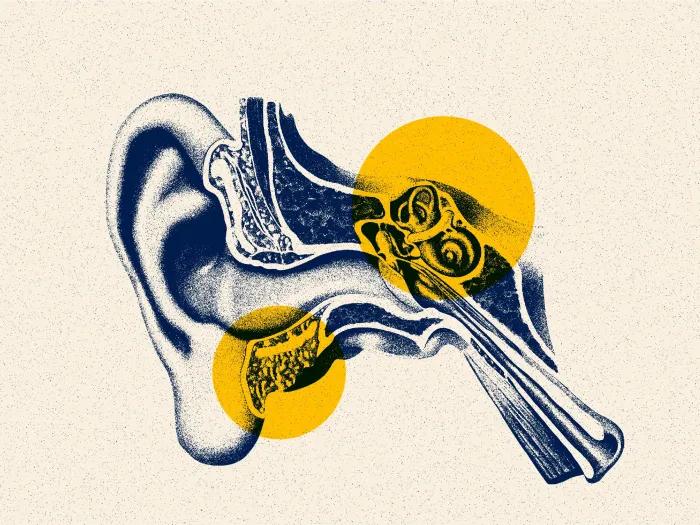
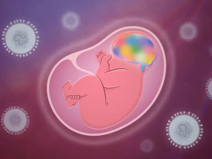
Microtia is a congenital anomaly characterized by the underdevelopment of the external ear (pinna) during the first trimester of gestation. Derived from the Latin terms for "small ear," the condition presents across a spectrum of anatomical variations, ranging from mild structural deficiencies to a small, vestigial remnant often described as a "lobular" or "peanut" deformity. Microtia occurs in approximately 1 in 5,000 live births. While it affects all populations, clinical data indicates significant variance in prevalence based on biological sex and ethnic background. Microtia primarily concerns the malformation of the external ear, but it is frequently comorbid with conditions affecting the middle ear and auditory canal:
- Aural Atresia: The complete absence of the external auditory canal
- Canal Stenosis: A condition where the ear canal is present but significantly narrowed
Our Approach
Following the neonatal diagnosis of microtia, a multidisciplinary approach is employed to evaluate systemic health and auditory function. The primary objective is to identify any associated congenital anomalies and establish a long-term care plan. Patients and families have several pathways for managing microtia, ranging from multi-stage biological reconstruction to prosthetic options or observation.
- Autologous Rib Cartilage Reconstruction: At Michigan Medicine, we utilize the Firmin technique, a streamlined two-stage protocol.
- Medpor (Porous Polyethylene) Reconstruction: This method utilizes a synthetic, pre-fabricated framework rather than harvested cartilage.
- Auricular Prosthetics: A prosthetic ear is custom-crafted by an anaplastologist to mirror the patient's unaffected ear.
- Conservative Management (The "Do Nothing" Option): Choosing not to undergo surgical intervention is a medically valid pathway.
Is my child a candidate for microtia treatment?
Children with microtia require specialized evaluation by an Audiologist. If additional dysmorphic features or multi-system abnormalities are present, a referral to a Medical Geneticist is recommended. In cases where microtia is a component of a broader craniofacial syndrome—such as Treacher Collins or Goldenhar Syndrome—care is coordinated through a dedicated craniofacial team.
Appointment Information
For appointments, please call 734-936-5730.
Please visit the Microtia Multi-Disciplinary Clinic page for more information.
What can my child expect with microtia treatment?
The initial clinical evaluations involve:
- Renal Ultrasonography: A renal ultrasound is often indicated to assess for anatomical irregularities (e.g., renal agenesis or ectopia).
- Comprehensive Audiological Assessment: Determines the degree of conductive hearing loss and assesses the functional status of the middle and inner ear.
- Genetic Consultation: To identify potential underlying syndromes or chromosomal variations.
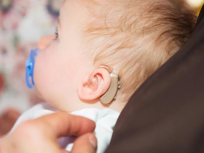
- Otolaryngology (ENT) Evaluation: To grade the severity of the microtia and confirm the presence or absence of the external auditory canal. The specialist will discuss options for surgical reconstruction and auditory habilitation, such as bone-conduction hearing aids.
For Autologous Rib Cartilage Reconstruction:
- Stage I: Rib cartilage is harvested and sculpted into a precise three-dimensional framework. This is implanted into a subcutaneous pocket. Suction drains are used to ensure the skin conforms to the new structure. This stage typically requires a 3 to 5-day hospital stay.
- Stage II: The ear is "released" or elevated from the scalp to provide natural projection. A skin graft is applied to the posterior (back) surface of the ear.
Risks & Benefits
Some of the risks include:
- Autologous Rib Cartilage Reconstruction: Donor Site Morbidity (Temporary pain at the rib harvest site and a chest scar); Unpredictability (Early healing of the graft can occasionally vary).
- Medpor (Porous Polyethylene) Reconstruction: The implant remains a foreign body and does not become "living" tissue. It lacks protective sensation (feeling), making it susceptible to unnoticed trauma. This carries a lifelong risk of extrusion (the implant pushing through the skin) or infection.
Benefits
- Autologous Rib Cartilage Reconstruction: Biocompatibility (No risk of graft rejection); Durability (The ear is living tissue that heals, bleeds, and grows; it is resistant to trauma); Longevity (Designed to last a lifetime).
- Medpor (Porous Polyethylene) Reconstruction: Eliminates the need for a donor site/chest surgery and offers highly consistent cosmetic contours.
- Auricular Prosthetics: Provides an exceptional cosmetic match with a minimally invasive, single-stage procedure.
- Conservative Management: Increased rib cartilage volume in older children allows for more detailed sculpting, and the patient can participate more actively in the decision-making process.
Locations
-
Pediatric Otolaryngology Clinic | C. S. Mott Children's Hospital 1540 E Hospital Dr
Floor 2 Reception A
Ann Arbor, MI 48109-4227Get Directions
Doctors
Jeffrey Tan Gu, MD
Clinical Assistant Professor
Otolaryngology, Plastic Surgery-Head & Neck
Adam Van Horn, MD
Clinical Assistant Professor
Pediatric Otolaryngology, Otolaryngology
News & Stories
Research may help better predict outcomes in kids with congenital cytomegalovirus
Human ability to focus on specific sounds not found to originate in auditory nerve, brainstem
Uncovering the link between a common congenital viral infection and autism

Sleep apnea disparities in kids: Obesity may override impact of race, socioeconomics
Study shows promising treatment for tinnitus
