Spina Bifida (Pediatric)
Spina Bifida (Pediatric)
What is Spina Bifida?
Myelomeningocele, or Spina Bifida, occurs in approximately one in every thousand live births. The condition typically arises when there is a defect in the base of the spinal cord such that the neural elements protrude through an opening in the skin. Often, there is disruption of signal flow between the brain and nerves below the level of the spinal cord defect, resulting in difficulties with walking and standing and causing bowel and bladder problems. In addition, the flow of cerebrospinal fluid around the brain and spinal cord is often affected, resulting in hydrocephalus that requires a shunt operation.
Myelomeningocele (A Severe Form of Spina Bifida)
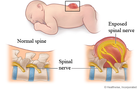
Myelomeningocele is a rare and severe form of spina bifida in which part of the spinal nerves or spinal cord is exposed on the outside of the skin. This type of spina bifida can lead to health problems, physical disability, and learning problems. Treatment usually includes surgery, braces or other corrective devices, physical therapy, and sometimes extra help in school.
Our Approach
The Myelomeningocele Clinic at U-M Health combines the expertise of Neurosurgery, Pediatrics, Orthopaedics, Urology, Physical Medicine & Rehabilitation, and other specialties to address the multiple medical needs of patients with these congenital spinal cord abnormalities. This multidisciplinary approach ensures that optimal and individualized care is provided to each patient in a regular and timely fashion.
For infants diagnosed with myelomeningocele prenatally, our multidisciplinary Fetal Diagnosis & Treatment Center offers expert management and fetal surgery when appropriate to treat myelomeningocele before a child is even born.
For adolescents with myelomeningocele, our multidisciplinary Physical Disabilities Transitional Care & Adult Clinic assists adolescent patients with the transition of medical care from pediatric to adult specialists. The clinic combines the expertise of pediatric and adult care specialists in urology, physical medicine and rehabilitation, gynecology and social work. Together, these seamlessly linked programs offer fully integrated care for families, ensuring individualized, coordinated care for children and adolescents with myelomeningocele and their families.
Appointment Information
For more information, call 734-936-5062.
What are the symptoms of spina bifida?
Your child's symptoms will depend on how severe the defect is. Most children with the mild form of spina bifida don't have any problems from it.
In many cases, children with meningocele don't have any symptoms.
Children with the most severe form of spina bifida often have spine and brain issues that cause serious problems. They may have:
- Little or no feeling in their legs, feet, or arms, so they may not be able to move those parts of the body.
- Bladder or bowel problems, such as leaking urine or having a hard time passing stools.
- Fluid buildup in the brain (hydrocephalus). Even when it is treated, this may cause seizures, learning problems, or vision problems.
- A curve in their spine, such as scoliosis.
How is spina bifida diagnosed?
During pregnancy, you can have a blood test (quad screen) and an ultrasound of the developing baby. These tests check for signs of spina bifida and other problems. If test results suggest a birth defect, you can choose to have an amniocentesis. This test helps confirm if the baby has spina bifida.
After birth, a doctor can usually tell if a baby has spina bifida by how the baby's back looks. If spina bifida is suspected, the doctor may do an X-ray, an MRI, or a CT scan to see if the defect is mild or severe.
How is spina bifida treated?
Most children with the mild form of spina bifida don't need treatment. Children with meningocele may not need treatment either. But children with the most severe form usually need surgery. Sometimes surgery to correct severe spina bifida can be done before a baby is born.
A child who has hydrocephalus will need surgery to put in a drainage tube called a shunt. It relieves pressure on the brain by draining excess fluid into the belly. This keeps the swelling from causing more damage to the brain.
Experts such as physical therapists and occupational therapists work with children who have severe spina bifida. The work starts soon after the child's birth. These therapists can teach parents and caregivers how to do exercises and activities with the child.
Some children may need a brace, a wheelchair, or other aids. Children with bladder control problems may need help using a catheter each day to prevent infection and kidney damage. To help prevent bowel problems, parents usually begin working with the doctor or nurse on managing bowel care as soon as the child starts eating solid food. As children with severe spina bifida grow, other treatments and surgeries may be needed to manage problems that arise.
Locations
-
Fetal Diagnosis and Treatment Center | Von Voigtlander Women's Hospital 1540 E Hospital Dr
Floor 9
Ann Arbor, MI 48109-4264Get Directions -
Pediatric Neurosurgery Clinic | C. S. Mott Children's Hospital 1540 E Hospital Dr
Floor 3 Reception B
Ann Arbor, MI 48109-4219Get Directions -
Pediatric Neurosurgery Clinic | Northville Health Center 39901 Traditions Dr
Floor 2
Northville, MI 48168-9493Get Directions -
Pediatric Neurosurgery Clinic | UM Health-Sparrow Professional Building 1200 E Michigan Avenue
Ste 340
Lansing, MI 48912Get Directions
Doctors

Hugh James Lauriston Garton, MD
Professor
Neurological Surgery

Erin Klements, MD
Clinical Instructor
Physical Medicine & Rehabilitation, Pediatrics, Pediatric Rehabilitation Medicine

Suresh Natesh Magge, MD
Clinical Professor
Neurological Surgery

Neena Ishwari Marupudi, MD
Clinical Associate Professor
Neurological Surgery

Karin Marie Muraszko, MD
Professor
Neurological Surgery
News & Stories

A concussion expert and football father discusses why he values teamwork over zero-risk
How a complex, 14-hour surgery saved a woman’s ‘hopeless’ spine
An AI model that can read and diagnose a brain MRI in seconds

Finally hope after 9 surgeries for tethered cord syndrome
Human ability to focus on specific sounds not found to originate in auditory nerve, brainstem