Retinoschisis
Retinoschisis
What is retinoschisis?
Retinoschisis is a condition in which an area of the retina (the tissue lining the inside of the back of the eye that transmits visual signals to the optic nerve and brain) has separated into two layers. The part of the retina that is affected by retinoschisis will have suboptimal vision. This can occur in different layers of the retina, and for different reasons.
The two major causes of retinoschisis are:
Juvenile X-linked Retinoschisis is a genetic disease of the retina and affects primarily boys and young men. In this form of retinoschisis, a mutation or abnormal gene is carried on the X chromosome. Men have only one X chromosome, while women have two. Therefore, women can carry the condition, but because they almost always have another normal X chromosome, they typically retain normal vision, even as carriers. Men, on the other hand, will develop subnormal vision if they have an affected X chromosome.
This condition typically presents during childhood and is estimated to affect one in 5,000 to 25,000 individuals. Affected males are usually identified in grade school, but occasionally are identified as young infants. Affected boys and men should have periodic eye examinations.
Degenerative retinoschisis is the splitting of the retina as a result of aging. It can affect men and women. This is not a genetic condition.
What are the risk factors of retinoschisis?
Retinal detachment can occur if the anchoring of the outer layer of the retina to the eye wall is impaired. Since retinoschisis patients are more susceptible to retinal detachment, they should have regular examinations with an ophthalmologist. When detected early, a complicating retinal detachment can be treated surgically; however, splitting or schisis of the retina itself cannot be corrected by medication or surgery.
Appointment Information
To schedule an appointment, please call 734-763-5874.
What are the symptoms of retinoschisis?
Some of the symptoms include decreased central vision and decreased peripheral vision. These symptoms may not necessarily mean that you have retinoschisis. However, if you experience one or more of these symptoms, contact your eye doctor for a complete exam.
How is retinoschisis diagnosed?
The electroretinogram (ERG) is used to assess function of the nerve tissue in the retina. The eye is stimulated with light after either dark or light adaptation. Contact lenses, embedded with an electrode to measure electrical impulses created by the functioning retina, are worn by the patient. The reaction of the eye to various light stimuli is recorded and evaluated. This test documents the type of photoreceptor activity and the overall function of the inner and outer layers of the retina, and is a very important tool in diagnosis.
ERGs are easily obtained on adults and children under 24 months of age. However, ERGs performed on children between the ages of two and five can be difficult and often require general anesthesia. Therefore, we encourage the testing be done before age two or after age five. In general, after age five, nearly all children can have an ERG without any difficulties.
Beginning at age 3-4, all children, even without retinoschisis, should have their vision checked yearly and glasses prescribed as necessary. Children who show schisis of the peripheral retina need more frequent examinations. We feel these examinations should be conducted by a subspecialty trained retina surgeon, since surgery would be required if a retinal detachment were to develop. Follow-up intervals are best determined by the doctor who is following your child.
How is retinoschisis treated?
Carbonic anhydrase inhibitors might be beneficial in reducing the cystic spaces observed in X-linked retinoschisis. There is no medical treatment for degenerative retinoschisis; however, vitrectomy surgery is occasionally required for complications related to either type of retinoschisis.
Glasses may improve the overall quality of vision in a patient with retinoschisis who is also near-sighted or farsighted, but will not "repair" the nerve tissue damage from the retinoschisis.
Vitamin A does not appear to help in retinoschisis. Vitamin A may have benefits for other genetic retinal diseases, particularly in cases of retinitis pigmentosa where the retinal nerve cells are slowly dying. However, retinoschisis is quite different from retinitis pigmentosa, since the retinal cells and their connections in retinoschisis are mechanically disrupted but are not thought to be dying.
Locations
-
Ophthalmology Clinic | Brighton Center for Specialty Care 7500 Challis Rd
Entrance 1, Level 2
Brighton, MI 48116-9416Get Directions -
Ophthalmology Clinic | Kellogg Eye Center-Grand Blanc 3181 E Grand Blanc Rd
Grand Blanc, MI 48439-2709Get Directions -
Retina & Uveitis Clinic | Kellogg Eye Center 1000 Wall St
Elevator B Floor 2
Ann Arbor, MI 48105-1912Get Directions
Doctors

Cagri Giray Besirli, MD, PhD
Associate Professor
Ophthalmology

Grant Michael Comer, MD, MS
Clinical Associate Professor
Ophthalmology

Emily Ann Eton, MD
Clinical Assistant Professor
Ophthalmology

Abigail Teich Fahim, MD, PhD
Associate Professor
Ophthalmology

Thomas Wright Gardner, MD, MS
Professor
Ophthalmology

Michael Jacob Huvard, MD
Clinical Assistant Professor
Ophthalmology

Mark W Johnson, MD
Professor
Ophthalmology

Shilpa Kodati, MBBS
Assistant Professor
Ophthalmology

Jason Matthew Lewis Miller, MD PhD
Assistant Professor
Ophthalmology

Rajesh Chalamalasetty Rao, MD
Associate Professor
Ophthalmology




News & Stories
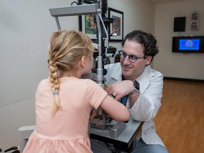
Innovative children’s vision research gets a boost from the Knights Templar Eye Foundation
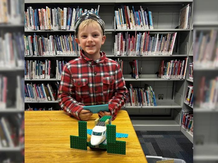
Nerf gun accident causes retinal detachment in pediatric patient

Kellogg care inspires gift to vision research

Mediterranean diet can slow progression of age-related macular degeneration
Patient with retinitis pigmentosa receives new hope at Kellogg’s low vision clinic