
Larynx (Voice Box) Cancer
Larynx (Voice Box) Cancer
Larynx Cancer (also called voice box cancer) have special importance because of their significant effects on voice, swallowing and quality of life. In the United States, it is estimated that over 12,000 new cases are diagnosed each year and that this incidence is increasing during a time that many other cancers are decreasing.
Larynx (voice box)
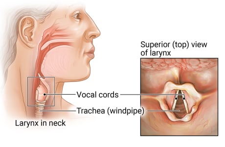
The voice box (larynx) is located in the neck, just above the windpipe (trachea). The two vocal cords in the larynx are covered with a mucous membrane. When air passes through the vocal cords, they vibrate and produce sound.
There are three main parts of the larynx:
- Supraglottis: The upper part of the a larynx; above the vocal cords.
- Glottis: The middle part of the larynx where the vocal cords are located.
- Subglottis: The lower part of the larynx between the vocal cords and windpipe (trachea).
Appointment Information
The Head & Neck Oncology Program offers diagnosis and treatment for those with laryngeal cancer. To make an appointment, please call 734-936-8051.
Healthcare professionals, please contact our M-LINE service: 800-962-3555.
What are the symptoms of laryngeal cancer
In general, the typical signs of laryngeal cancer include a sore throat and ear pain. These symptoms can be caused by other types of conditions and disease. Check with your doctor if you have any of the following:
- A sore throat or cough that does not go away
- Trouble or pain when swallowing
- Ear pain
- A lump in the neck or throat
- A change or hoarseness in the voice
How is laryngeal cancer diagnosed?
If your doctor suspects laryngeal cancer, they will do the following:
- Physical exam of the throat and neck
- Biopsy: The removal of cells or tissues for viewing under a microscope by check for signs of cancer. There are three types of biopsy:
- Fine-needle aspiration: This is the removal of tissue or fluid using a needle.
- Incisional biopsy: The removal of part of tissue that looks abnormal.
- Excisional biopsy: The entire removal of tissue that looks abnormal.
- Laryngoscopy: This procedure checks the voice box (also called the larynx) with a mirror or a laryngoscope (a thin, tube-like instrument with a light and a lens for viewing).
- Endoscopy: This procedure allows the doctor to view inside the throat, esophagus and trachea to check for abnormal areas. An endoscope (a thin tube with a light and lens for viewing) is inserted into the mouth. Usually there's a tool at the end of the endoscope that allows the doctor to collect a sample of abnormal tissue.
- An MRI: A procedure that uses a magnet, radio waves, and a computer to make a series of detailed pictures of areas inside the body. This procedure is also called nuclear magnetic resonance imaging (NMRI).
- CT scan (CAT scan): This procedure is also called computed tomography, computerized tomography, or computerized axial tomography. Pictures are taken of areas inside the body using a computer linked to an x-ray. In some cases, a dye may be injected or swallowed in order for the organs and tissues to be seen more clearly.
- PET-CT scan: This procedure combines the pictures from a positron emission tomography (PET) scan and a CT scan. These scans are done at the same time using the same machine. The combination provides more detailed images of the inside of the throat and voice box.
- Bone scan: A very small amount of radioactive material is injected into the vein. It collects in the bones and a scanner can then detect signs of cancer in the bones.
- Barium swallow: The patient drinks a liquid containing barium (a silver-white metallic compound) that coats the esophagus and stomach. After drinking barium, X-rays are taken and provide another way to view abnormal tissue.
How is laryngeal cancer treated?
The Head and Neck Oncology Program provides treatment which includes surgery or radiation with or without chemotherapy. A number of complex factors go into our treatment recommendations. Depending on the size and type of tumor, we may perform a partial laryngectomy, often via robotic-assisted surgery. Robotic surgery allows us to more easily reach the tumor and avoid placing a temporary breathing tube in the neck. A partial laryngectomy lets us remove smaller tumors while preserving your voice.
Radiation may provide an option for early-stage voice box cancer. For more advanced cancers, a combination of radiation therapy and chemotherapy can offer an alternative to surgery while preserving the voice box. In some cases, we may recommend complete removal of the voice box, also called a total laryngectomy. Learn more about radiation therapy, please visit our Radiation Therapy webpage.
Locations
-
Otolaryngology Clinic | Northville Health Center 39901 Traditions Dr
Floor 2
Northville, MI 48168-9493Get Directions -
Otolaryngology Clinic | Taubman Center 1500 E Medical Center Dr
Floor 1 Reception A
Ann Arbor, MI 48109-5312Get Directions
Doctors

Marisa Rae Buchakjian, MD, PhD
Clinical Assistant Professor
Otolaryngology

Keith Andrew Casper, MD
Clinical Associate Professor
Otolaryngology

David WA Forner, MD, MSc, FRCSC
Clinical Assistant Professor
Otolaryngology

Molly Elaine Heft Neal, MD
Clinical Assistant Professor
Otolaryngology

Kelly Michele Malloy, MD
Clinical Professor
Otolaryngology

Scott Alan Mclean, MD, PhD
Clinical Associate Professor
Otolaryngology, Facial Plastic Surgery

Mark Edward P Prince, MD
Professor
Otolaryngology

Andrew Gregg Shuman, MD
Professor
Otolaryngology

Chaz Luke Stucken, MD
Clinical Associate Professor
Facial Plastic Surgery, Otolaryngology, Plastic Surgery-Head & Neck

Pratyusha Yalamanchi, MD, MBA
Clinical Assistant Professor
Otolaryngology
