Dynamic Airway Collapse
Dynamic Airway Collapse
What is dynamic airway collapse?
The upper airway is what connects the back of the throat down to the individual lungs. This is composed of the larynx (voice box), trachea (windpipe), and bronchi (branches of windpipe connecting to the lungs). Tracheobronchomalacia (TBM) is a condition in which the cartilage in the front walls of the airway softens resulting in a floppy or weak airway that collapses with breathing and makes you short of breath. Excessive dynamic airway collapse (EDAC) refers to when the airways collapse due to bulging of the back, soft part of the airway.
What causes dynamic airway collapse?
Tracheobronchomalacia has multiple causes. Infants may be born with the disorder, or adults may develop it later on in life. The most common causes of tracheobronchomalacia include:
- Severe persistent asthma
- Chronic infections (such as bronchitis)
- Emphysema
- Gastroesophageal reflux disease (GERD)
- Chronic use of steroids
- Excessive weight gain
- Damage to the trachea or esophagus caused by surgery or other medical procedures
- Damage caused by a long-term breathing tube or tracheostomy
- Inhaling irritants
- Polychondritis (inflammation of cartilage in the trachea)
Appointment Information
To make an appointment, call 734-936-8051.
What are the symptoms of dynamic airway collapse?
The symptoms of tracheobronchomalacia or excessive dynamic airway collapse include:
- Abnormal/Irregular breathing noises (such as rattling sounds)
- Chronic cough
- Difficulty swallowing, especially solid foods
- Recurring respiratory tract infections
- Shortness of breath
How is airway stenosis diagnosed?
Your evaluation is customized to your condition. Your evaluation may include:
- A complete history of your breathing problem
- A physical examination of your head, neck, and chest
- Evaluation your breathing ability through pulmonary function testing (PFTs), and a 6 minute walk test
- Examination of your throat, voice box, and airway through endoscopy in the clinic or operating room
- Imaging of your airways via X-rays or computed tomography (CT) scans. This may include dynamic 3D chest CT scans, a non-invasive procedure performed with the patient inhaling and exhaling to examine how the airways change with breathing.
How is dynamic airway collapse treated?
Your doctor may recommend non-surgical or surgical treatment options depending on your symptoms and other factors.
Non-surgical treatment
In many cases, tracheobronchomalacia or excessive dynamic airway collapse gradually improves with improvement in contributing medical conditions. However, patients who suffer from frequent respiratory infections or persistent symptoms should be closely monitored. Their options for treatment may include the following:
- Optimizing medically the conditions that could be exacerbating tracheobronchomalacia.
- Chest physical therapy: non-invasive techniques that include lightly tapping on the chest to break up mucus, and deep breathing exercises.
Continuous positive airway pressure (CPAP): through a face mask, air under mild pressure forces the trachea to remain open during breathing.
Surgery
For patients who do not benefit from conservative treatment, we offer surgical procedures to improve symptoms with tracheobronchomalacia or excessive dynamic airway collapse. These include:
- Tracheobronchial stenting: while examining your airway through the mouth while you are asleep, the airway is propped open with a thin metallic or silicone expandable stent placed into the airway through a bronchoscope. Most stents are placed as a trial to evaluate if splinting of the airway will improve symptoms before definitive treatment.
- Tracheal resection and reconstruction: via an incision through the neck or chest, the floppy area of the airway is cut away (resected), and the two remaining ends of the airway are sewn back together, resulting in an unobstructed airway. This procedure is best for patients with a small area of their airway affected.
- Endoscopic laser tracheobronchoplasty: while examining your airway through the mouth while you are asleep, a laser is used through a flexible scope to stiffen the tissues that collapse during breathing to improve the airway staying open.
- Open tracheobronchoplasty: a surgical procedure that provides support to the weak/floppy airway, preventing its collapse during breathing. Via an incision through the neck or chest, a mesh or surrounding tissue is placed as support, and the length of the floppy airway is sutured to it so that the airway is held in an open configuration during the breathing cycle.
- Tracheostomy: an incision is made from the front of the neck into the airway, and a tube is inserted into the airway to hold open the area of collapse and allow normal breathing.
Locations
-
Otolaryngology Clinic | Brighton Center for Specialty Care 7500 Challis Rd
Entrance 1, Level 2
Brighton, MI 48116-9416Get Directions -
Otolaryngology Clinic | West Ann Arbor Health Center-Parkland Plaza 380 Parkland Plaza Ste 210
Floor 2
Ann Arbor, MI 48103-6201Get Directions -
Vocal Health Center | Taubman Center 1500 E Medical Center Dr
Floor 1 Reception A
Ann Arbor, MI 48109-5312Get Directions
Doctors

Norman Dertad Hogikyan, MD, F.A.C.S.
Professor
Otolaryngology

Robbi Ann Kupfer, MD
Clinical Associate Professor
Otolaryngology

Robert James Morrison, MD
Clinical Associate Professor
Otolaryngology
News & Stories

Rural-urban disparities in head and neck cancer

Biomarkers aid cost-benefit analysis of head and neck cancer immunotherapy

Gratitude inspires gift to improve survivorship care
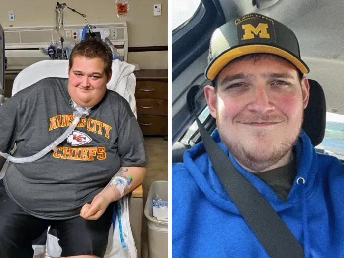
Weight loss and surgery restore patient’s throat after near-death brush with likely COVID
A link between e-cigarettes and oral cancer