Diabetic Retinopathy
Diabetic Retinopathy
What is diabetic retinopathy?
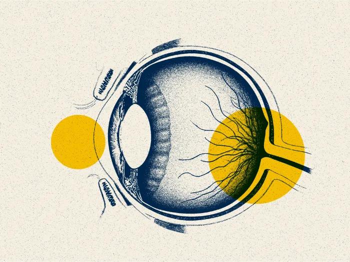
Diabetic retinopathy is caused by changes in the blood vessels of the retina. When these blood vessels are damaged, they may leak blood and grow fragile new vessels. When the nerve cells are damaged, vision is impaired. These changes can result in blurring of your vision, hemorrhage into your eye, or, if untreated, retinal detachment. Diabetic retinopathy is the most common diabetic eye disease and a leading cause of blindness in the U.S.
What causes diabetic retinopathy?
People with untreated diabetes are 25 times more at risk for blindness than the general population. The longer a person has had diabetes, the higher the risk of developing diabetic retinopathy. Fortunately, with regular, proper eye care and treatment when necessary, the incidence of severe vision loss has been greatly reduced. If you have diabetes, your ophthalmologist can help to prevent serious vision problems.
Diabetic retinopathy can cause vision loss in two ways:
- Macular Edema: Macular edema is a condition where your retinal blood vessels develop tiny leaks. When this occurs, blood and fluid leak from the retinal blood vessels and fatty material (called exudate) is deposited in the retina. This causes swelling of the retina and is called diabetic macular edema. When this swelling occurs in the central part of the retina, also known as the macula, your vision will be reduced or blurred.
- Proliferative Retinopathy and Vitreous Hemorrhage: Proliferative retinopathy refers to the changes that occur when new, abnormal blood vessels begin to grow on the surface of your retina. This abnormal growth is called neovascularization. If these abnormal blood vessels grow around your pupil, glaucoma can result from the increasing pressure within your eye. These new blood vessels have weaker walls and may break and bleed, or cause scar tissue to grow that can pull the retina away from the back of your eye. When your retina is pulled away it is called a retinal detachment and, if left untreated, it can cause severe vision loss, including blindness. Leaking blood can cloud the vitreous—the clear, jelly-like substance that fills the back of the eye—and block the light passing through the pupil to the retina, causing blurred and distorted images. In more advanced proliferative retinopathy, diabetic fibrous or scar tissue can form on the retina.
What are the risk factors of diabetic retinopathy?
- Poorly-controlled diabetes
- High blood pressure
- Long-term duration of diabetes
- Elevated blood cholesterol levels
- Sleep apnea
Appointment Information
To schedule an appointment, please call 734-764-4190.
What are the symptoms of diabetic retinopathy?
- Blurred vision
- Sudden loss of vision in one eye
- Seeing rings around lights
- Dark spots or flashing lights
The symptoms described above may not necessarily mean that you have diabetic retinopathy. However, if you experience one or more of these symptoms, contact your ophthalmologist for a complete exam. It is also important to note that pregnancy and high blood pressure may aggravate diabetic retinopathy.
How is diabetic retinopathy diagnosed?
Macular edema and proliferative diabetic retinopathy can be assessed through a dilated eye exam. In addition, tests such as a fluorescein angiogram and ocular coherence tomography (OCT) can be conducted. The angiogram test involves the injection of a contrast agent or dye into your arm. The contrast agent can then be seen coursing through the blood vessels in your retina. Normal, healthy blood vessels do no leak. However, damaged blood vessels in patients with macular edema will leak. In addition, the new vessels that develop in proliferative diabetic retinopathy also will leak the contrast agent. In this way, the damaged vessels and abnormal new vessels are identified.
Macular edema also can be assessed by using an OCT. This test uses light in a manner similar to ultrasound. The light is reflected back from the different layers of the retina and a cross sectional image of the retina is produced. Macula edema is noted when areas of your retina are shown to contain spaces filled with fluid.
How is diabetic retinopathy treated?
In mild cases, treatment for diabetic retinopathy is not necessary. Regular eye exams are critical for monitoring progression of the disease. Strict control of blood sugar and blood pressure levels can greatly reduce or prevent diabetic retinopathy. In more advanced cases, treatment is recommended to stop the damage of diabetic retinopathy, prevent vision loss, and potentially restore vision.
Treatment options include:
- Anti-VEGF therapy (Avastin, Lucentis, Eylea): Anti-VEGF therapy involves the injection of the medication into the back of your eye. The medication is an antibody designed to bind to and remove the excess VEGF (vascular endothelial growth factor) present in the eye that is causing the disease state. The FDA has approved Lucentis for macular edema and additional treatment options include Avastin and Eylea.
- Intraocular steroid injection: Intraocular steroid injection is a treatment for diabetic macular edema. This therapy helps reduce the amount of fluid leaking into your retina, resulting in visual improvement. Due to the chronic nature of diabetic eye disease, this treatment may need to be repeated or combined with laser therapy to obtain maximum or lasting effect.
- Laser surgery: Laser surgery is often helpful in treating diabetic retinopathy. To reduce macular edema, a laser is focused on the damaged retina to seal leaking retinal vessels. For abnormal blood vessel growth (neovascularization), the laser treatments are delivered over the peripheral retina. The small laser scars that result will reduce abnormal blood vessel growth and help bond the retina to the back of your eye, thus preventing retinal detachment. Laser surgery may be performed in your ophthalmologist's office or in an outpatient clinic. Laser surgery can greatly reduce the chance of severe visual impairment.
- Vitrectomy: Vitrectomy may be recommended in advanced proliferative diabetic retinopathy. During this microsurgical procedure that is performed in the operating room, the blood-filled vitreous is removed and replaced with a clear solution. Your ophthalmologist may wait several months to a year to see if the blood will clear on its own before going ahead with surgery. In addition to a vitrectomy, retinal repair may be necessary if scar tissue has detached the retina from the back of your eye. Severe loss of vision or even blindness can result if surgery is not performed to reattach the retina.
Patient Resources
Watch these video animations to learn more about diabetic retinopathy, the affect that the diabetic retinopathy has on the eyes, and tests and treatments options for the condition.
Topics covered
- Vitreous Hemorrhage
- Macular Edema and Macular Ischemia
- Non-proliferative Diabetic Retinopathy (NPDR)
- Proliferative Retinopathy (PDR)
- Fluorescein Angiography Test
- Ocular Coherence Tomography (OCT) Test
- Ultrasound Test
- Anti-VEGF therapy (Avastin, Lucentis, Eylea) Treatment
- Laser Surgery Treatment
- Vitrectomy Treatment
- Panretinal Photocoagulation (PRP) Treatment
- Diabetic and Your Eyes
Locations
-
Ophthalmology Clinic | Brighton Center for Specialty Care 7500 Challis Rd
Entrance 1, Level 2
Brighton, MI 48116-9416Get Directions -
Ophthalmology Clinic | Kellogg Eye Center-Grand Blanc 3181 E Grand Blanc Rd
Grand Blanc, MI 48439-2709Get Directions -
Retina & Uveitis Clinic | Kellogg Eye Center 1000 Wall St
Elevator B Floor 2
Ann Arbor, MI 48105-1912Get Directions
Doctors

Cagri Giray Besirli, MD, PhD
Associate Professor
Ophthalmology

Grant Michael Comer, MD, MS
Clinical Associate Professor
Ophthalmology

Emily Ann Eton, MD
Clinical Assistant Professor
Ophthalmology

Abigail Teich Fahim, MD, PhD
Assistant Professor
Ophthalmology

Thomas Wright Gardner, MD, MS
Professor
Ophthalmology

Michael Jacob Huvard, MD
Clinical Assistant Professor
Ophthalmology

Mark W Johnson, MD
Professor
Ophthalmology

Shilpa Kodati, MBBS
Assistant Professor
Ophthalmology

Jason Matthew Lewis Miller, MD PhD
Assistant Professor
Ophthalmology

Rajesh Chalamalasetty Rao, MD
Associate Professor
Ophthalmology


News & Stories

Using genetic testing, doctors help patient find answers for her diabetes
A new clue for aging eyes

Supplementing with peptides: Good for extra pep or a needless step?

Susan J. Lane: Gratitude and Giving
A unique patient case inspiring research