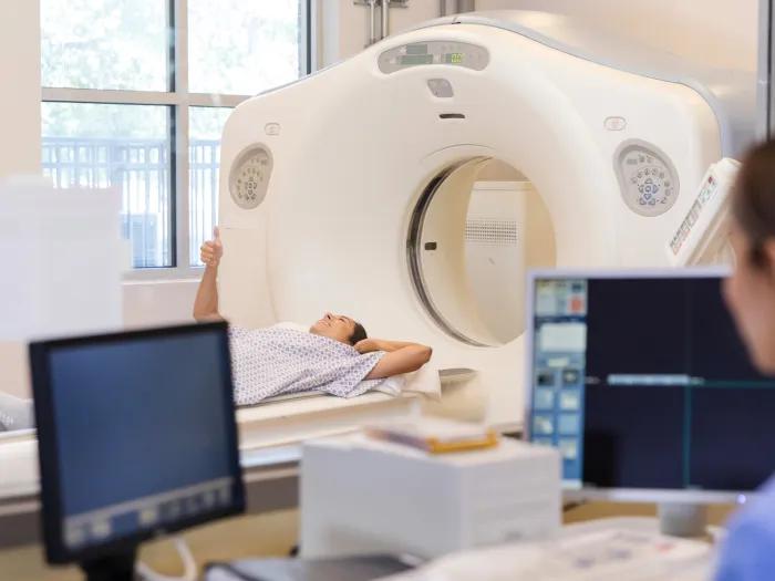
Computed Tomography (CT scan)
Computed Tomography (CT scan)
What is computed tomography (CT scan)?
CT scanning, also called computed or computerized tomography, is an x-ray test used for diagnosis. X-rays are taken from a series of different angles and arranged by a computer to show a cross-sectional view of organs in the body.
Our Approach
The U-M Health Radiology offers standard CT Studies as well as cutting-edge high resolution and 3D CT angiography, all by board-certified, fellowship-trained experts.
Our divisions include:
- Abdominal - examinations of the abdomen and pelvis for detection of the full spectrum of conditions, plus advanced techniques, such as evaluation of abdominal aortic aneurysms for stent graft placement
- Cardiothoracic – we perform more than 8,000 chest CT exams annually, including CT pulmonary angiography for the diagnosis of acute and chronic pulmonary embolism
- Musculoskeletal – full range of bone, joint and tissue CT scanning, including CT arthrography, which detects tears and their extent, such as for rotator cuffs
- Neuroradiology – a full spectrum of diagnostic and research CT applications for the nervous system, spine, and the head and neck
Lowest Dose of Radiation
Our studies and protocols make sure our patients receive the lowest dose of radiation required for their study. Having the very latest equipment allows for studies to be done quicker, which also lowers radiation exposure.
We utilize the General Electric Discovery CT750 HD, which provides up to a 50% lower dose of radiation for our patients, along with high-definition image quality for any part of the body.
3D CT Angiography
We also offer 3D CT angiography (to view major blood vessels) through our 3D Imaging Laboratory. We can use 3D technology to detect artery blockages, cerebral aneurysms and other vascular diseases, plus aid in pre-operative analysis and planning.
News & Stories
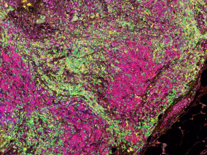
New mouse model for liposarcoma can help uncover new therapies

An AI model that can read and diagnose a brain MRI in seconds
Hospital partnership improves follow up scans, decreases long term risk after aortic repair
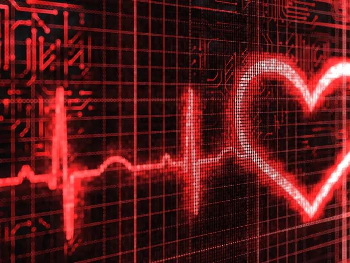
AI model helps diagnose often undetected heart disease from simple EKG

Understanding Prostate Biopsy