Ureteroscopy
Ureteroscopy
What is ureteroscopy?
Ureteroscopy is a minimally invasive method to treat kidney stones as well as stones located in the ureter. It is performed in the operating room with general or spinal anesthesia and is typically an outpatient procedure (you go home the same day).
Why is ureteroscopy done?
Urologists use ureteroscopy to remove stones that are stuck in the ureter and are closer to the bladder than the kidney (in the lower third of the ureter). But newer technology is allowing ureteroscopy to be used even for small stones in or near the kidney.
Appointment Information
U-M Health Urology is accepting new patients for initial and second opinions. Our medical staff’s goal is to see and evaluate patients based on their symptoms and urologic needs in a timely manner. For more information, call 734–936–7030.
How is ureteroscopy done?
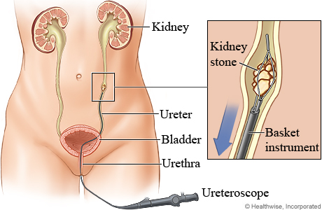
During ureteroscopy, the doctor passes a thin viewing instrument (ureteroscope) through your urethra and bladder into your ureter. The doctor moves the scope through your ureter until it reaches the location of the kidney stone. No cuts are made in the body.
Your doctor can take out the kidney stone using a small "basket" that comes out of the end of the ureteroscope. Small stones can be removed all in one piece. Larger stones may need to be broken up before the doctor can remove them.
How do I care for myself after ureteroscopy?
Most people are able to go home the same day of the procedure. But you may need to stay in the hospital. If you do, the stay is usually no more than 24 to 48 hours.
For several hours after the procedure you may have a burning feeling when you urinate. This feeling should go away within a day. Drinking a lot of water can help. Your doctor also may advise you to take medicine that numbs the burning. This medicine is called phenazopyridine. It is available by prescription and over the counter. Brand names include Pyridium and Uristat.
You may have some blood in your urine for 2 or 3 days.
Your doctor may prescribe an antibiotic for a day or two. This will help prevent an infection.
Doctors
Sapan Nitin Ambani, MD
Clinical Associate Professor
Urology
Casey Andrew Dauw, MD
Clinical Associate Professor
Urology

Khurshid Ridwan Ghani, MBCHB
Clinical Professor
Urology
William Woodruff Roberts, MD
Professor
Urology
News & Stories

Researchers create new path to target hard-to-drug prostate cancer protein
Urine-based test detects aggressive prostate cancer
How donor eggs helped one couple build the family they always hoped for
What causes infertility? A doctor answers common questions and clears up misconceptions

Insurance that covers male infertility procedures improves opportunities for family building