How Is ALS Diagnosed?
Diagnosing Amyotrophic Lateral Sclerosis (ALS) can be difficult because there is no single test or procedure to confirm the disease. Moreover, since many neurologic diseases cause similar symptoms, appropriate tests must be conducted to exclude the possibility of other conditions first.
Diagnosing ALS may involve:
- Electrodiagnostic tests (EMG/NCS): Electomyography (EMG) and nerve conduction study (NCS) evaluates muscle and nerve functions. Specific abnormalities in the NCS and EMG may suggest, for example, that the individual has a form of peripheral neuropathy (damage to peripheral nerves) or myopathy (muscle disease) rather than ALS.
- Magnetic resonance imaging (MRI): A noninvasive procedure that uses a magnetic field and radio waves to take detailed images of the brain and spinal cord.
How Is ALS Treated?
Although there is no known cure for ALS, treatment may help relieve symptoms and improve the quality of life. Treatments may include:
Medications
We at Michigan Medicine prescribe medications that are approved for ALS including Riluzole and Edaravone.
- Riluzole: Clinical trials with ALS patients showed that Riluzole prolongs survival by several months, mainly in those with difficulty swallowing. The drug also extends the time before an individual needs ventilation support.
- Edaravone: The medicine works by relieving the effects of oxidative stress, which may be related to the death of motor neurons (nerve cells) in people with ALS. Keeping motor neurons healthy may help to preserve muscle function.
- Relyvrio
- Tofersen
Physicians can also prescribe medications to help reduce fatigue, ease muscle cramps, and reduce excess saliva and phlegm. Drugs also are available to help patients with pain, depression, sleep disturbances, and constipation.
Therapies
- Physical and occupational therapy: Physical therapy and special equipment can enhance an individual’s independence and safety throughout the course of ALS. Gentle, low-impact aerobic exercise such as walking, swimming, and stationary bicycling can strengthen unaffected muscles, improve cardiovascular health, and help patients fight fatigue and depression.
- Speech therapy: As ALS progresses, speech therapists can help people develop ways for responding to yes-or-no questions with their eyes or by other nonverbal means and can recommend aids such as speech synthesizers and computer-based communication systems. These methods and devices help people communicate when they can no longer speak or produce vocal sounds.
- Breathing devices: When the muscles that assist in breathing weaken, use of nocturnal ventilatory assistance may be used to aid breathing during sleep. These devices may be used full-time as the disease reaches more advanced stage.
- Feeding tube: When individuals can no longer get enough nourishment from eating, doctors may advise inserting a feeding tube into the stomach. The use of a feeding tube also reduces the risk of choking and pneumonia that can result from inhaling liquids into the lungs.
What is a feeding tube?
A feeding tube is a small, flexible tube, about the diameter of a pencil or smaller, used to allow liquid nourishment to enter the stomach directly, bypassing the mouth, throat and esophagus. The feeding tube is often called a PEG tube, which stands for percutaneous (through the skin) endoscopic (using a scope) gastrostomy (an opening in the stomach) tube. It is also called a gastrostomy tube, or G-tube.
Why consider a feeding tube?
Some people that have ALS can develop difficulty swallowing, and this can develop significantly over time. This can lead to malnutrition and weight loss from inadequate protein and calorie intake. When this happens, the body uses muscle as a source of energy and protein and this accelerates the progression of weakness.
A PEG tube or feeding tube can restore adequate protein and calorie intake, and helps people feel better. The risk of swallowing difficulty is that food or liquid taken through the mouth may be inhaled into the airways rather than the stomach. This is called aspiration, and causes infection and pneumonia. Thin liquids and foods that crumble are especially likely to be aspirated.
When is the right time to put in a feeding tube?
Because of the nutritional deficiencies and the risk of aspiration, we recommend placing a PEG tube relatively soon after swallowing symptoms begin. One of the reasons is that progression of breathing muscle weakness often occurs at the same time swallowing weakness does. When breathing is stronger, placement of these tubes is easier and safer. Also, good nutrition and hydration can help to preserve available breathing muscle strength for as long as possible.
Can you still eat and drink if you have a PEG tube?
The gastrostomy tube is an alternative way of eating and drinking, but this does not affect eating by mouth. In the earlier stages of swallowing weakness, the tube is used to supplement oral intake. But if the weakness progresses and swallowing foods and fluids becomes too much work or becomes dangerous, it becomes the sole method of nutrition and hydration. There are many liquid formulations that give a 100% complete and balanced diet. If someone is still able to safely swallow small amounts of foods for pleasure, it is appropriate to do so.
Can medication be given through a PEG?
Many medications can be crushed, dissolved and given through the PEG. Some capsules can be opened and sprinkled into water or liquid food. Medication taken this way should be well mixed with water so that particles do not remain in the tube, where they may solidify and cause a blockage.
No time-released medications may be crushed or dissolved to administer through the PEG. If there is a liquid preparation of a medication, it can be easily given through the tube. Some medications come in patches and are absorbed through the skin. These include certain hormone supplements and some blood pressure medicines. Check with your doctor or pharmacist about alternative preparations of your medications.
What about heartburn?
If reflux (movement of stomach acid from the stomach up to the esophagus or throat) is a problem, medications to reduce acid formation are useful. This is not caused by ALS, but inflammation of the stomach or esophagus is common and can result in stomach pain or heartburn when using the PEG. If someone experiences reflux of food up the esophagus, a different medication or feeding pattern is recommended.
What is involved in placing a PEG tube?
The actual placement of the tube generally takes about a half hour. It is done with sedation given in a vein, and numbing medication at the insertion site on the stomach. Most people experience moderate pain for a few days after the tube in placed, and it is usually relieved well with over-the-counter pain medications.
The day of the procedure, a nutritionist meets with you and calculates your nutritional needs and indicates to the physician the type and amount of formula to prescribe. Food and supplies will be delivered to your home. You will also be taught how often to feed, the length of time for each feeding and how much free water to take by tube each day. A nurse will come to your home the day after your procedure to start your feedings and teach you and your family how everything works.
Your feeding may be given manually, from a bag that empties by gravity, or by a pump, which delivers a specific amount of the feeding formula per hour. You may discuss both alternatives during your visit with the Gastroenterologist (the digestive system doctor) before the placement of your tube. You may also discuss the type of feeding schedule that will best meet your needs. Some or all of your feeding can be given during the night while you sleep if that works best with your schedule and your particular digestive tract.
It is important that you begin to feed slowly in the beginning to avoid gas, bloating and/or diarrhea by going too fast. Over time, the number of feedings per day will be reduced, and the volume and speed of feedings increased as your stomach gets used to this new food.
Is a PEG tube noticeable?
The PEG tube is underneath your clothing and not visible when not in use. It can be formed into a loop and taped flat against the stomach. It is very easily concealed.
What happens if the PEG tube accidentally comes out?
If the tube is accidentally pulled out, it can usually be replaced easily at an emergency room with a Foley catheter, which is used for bladder drainage. The Foley has a bulb at the tip that is inflated after it is inserted to hold it in place. This is easy to do, and no anesthesia or sedation is needed. However, it should be done within hours of the PEG coming out, because if you wait until the next day, the small hole from the skin to the stomach may already be sealing up. With the site secured open with the Foley, your stomach doctor can then place a new PEG easily.
How do most people like the PEG tube?
We have not had patient regret getting PEG tubes. PEG tubes help people to live well, remain well nourished, and well hydrated. They greatly reduce the work of eating, and weight loss can be stopped or reversed. Some people can return to their usual body weight after insertion.
All our muscles and organs require good energy from good nutrition, including our breathing muscles. PEG tubes can help to improve quality and length of life. Good nutrition and hydration gives your body what it needs to fight the disease, repair damage and to try to grow new nerves. It is the best fight anyone has against ALS.
Researching the Future of ALS
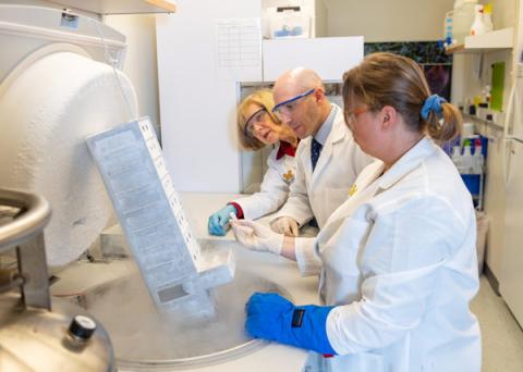
Michigan Medicine offers cutting edge research and every patient that comes to our clinic can participate in some form of research. While treating persons with ALS or other motor neuron diseases is a large part of our clinic, we are also at the forefront of ALS research.
Using many approaches, including basic laboratory research, epidemiologic research, and clinical trials, the University of Michigan is making major contributions to our understanding of ALS and the discovery of new treatment options.
One major research focus is identifying why people living in Michigan seem to be at a higher risk of ALS compared to other states. Led by Dr. Stephen Goutman, we evaluate for environmental risk factors via a survey and house an ALS patient biorepository that provides samples of biofluids (such as blood) to ALS researchers.
Researchers under the direction of Dr. Eva Feldman use these biofluids to better understand changes in gene expression and the immune system. These biospecimens are used in the laboratory to understand ALS pathogenesis and create models to screen and identify new therapies.
Clinical Trials
- We recently completed a safety study of stem cell therapy for the treatment of ALS led by Dr. Eva Feldman.
- We are active with the Northeast ALS Consortium (NEALS) and have recently participated in studies of retigabine, mexiletine, and tirasemtiv. Our current trials can be found at ClinicalTrials.gov
Make an Appointment
To request an appointment or to get more information, please call 734-936-9006 and a team member will get back to you within two business days.
Online resources from the following organizations were cited for information on Amyotrophic lateral sclerosis (ALS), symptoms, diagnosis and treatment:
- National Institute of Neurological Disorders and Stroke
- National ALS Registry


