Magnetic Resonance Imaging (MRI)
Test Overview
Magnetic resonance imaging (MRI) is a test that uses a magnetic field and pulses of radio wave energy to make pictures of organs and structures inside the body. In many cases, MRI gives different information about structures in the body than can be seen with an X-ray, ultrasound, or computed tomography (CT) scan. MRI also may show problems that cannot be seen with other imaging methods.
For an MRI test, the area of the body being studied is placed inside a special machine that contains a strong magnet. Pictures from an MRI scan are digital images that can be saved and stored on a computer for more study. The images also can be reviewed remotely, such as in a clinic or an operating room. In some cases, contrast material may be used during the MRI scan to show certain structures more clearly.
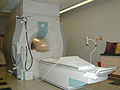
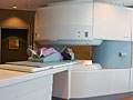
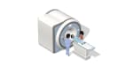
There are two main types of MRI—the standard MRI machine and the open MRI machine.
Why It Is Done
Magnetic resonance imaging (MRI) is done for many reasons. It is used to find problems such as tumors, bleeding, injury, blood vessel diseases, or infection. MRI also may be done to provide more information about a problem seen on an X-ray, ultrasound scan, or CT scan. Contrast material may be used during MRI to show abnormal tissue more clearly. An MRI scan can be done for the:
- Head.
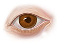
MRI can look at the brain for tumors, an aneurysm, bleeding in the brain, nerve injury, and other problems, such as damage caused by a stroke. MRI can also find problems of the eyes and optic nerves, and the ears and auditory nerves.
- Chest.
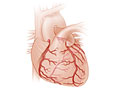
MRI of the chest can look at the heart, the valves, and coronary blood vessels. It can show if the heart or lungs are damaged. MRI of the chest may also be used to look for breast cancer.
- Blood vessels.
Using MRI to look at blood vessels and the flow of blood through them is called magnetic resonance angiography (MRA). It can find problems of the arteries and veins, such as an aneurysm, a blocked blood vessel, or the torn lining of a blood vessel (dissection). Sometimes contrast material is used to see the blood vessels more clearly.
- Abdomen and pelvis.
MRI can find problems in the organs and structures in the belly, such as the liver, gallbladder, pancreas, kidneys, and bladder. It is used to find tumors, bleeding, infection, and blockage. In women, it can look at the uterus and ovaries. In men, it looks at the prostate.
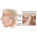
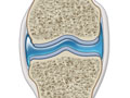
- Bones and joints.
MRI can check for problems of the bones and joints, such as arthritis, problems with the temporomandibular joint, bone marrow problems, bone tumors, cartilage problems, torn ligaments or tendons, or infection. MRI may also be used to tell if a bone is broken when X-ray results are not clear. MRI is done more commonly than other tests to check for some bone and joint problems.
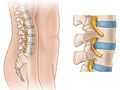
- Spine.
- MRI can check the discs and nerves of the spine for conditions such as spinal stenosis, disc bulges, and spinal tumors.
How To Prepare
For some MRI pictures of the belly, you may be asked to not eat or drink for several hours before the test.
Tell your doctor if you get nervous in tight spaces. You may get a medicine to help you relax. If you think you'll get this medicine, be sure to arrange a ride home. It may be unsafe for you to drive or get home on your own.
How It Is Done
Before the test
You will need to remove all metal objects (such as hearing aids, dentures, jewelry, watches, and hairpins) from your body. These objects may be attracted to the powerful magnet used for the test.
You will need to take off all or most of your clothes, depending on which area is examined. (You may be allowed to keep on your underwear if it's not in the way.) You will be given a gown to use during the test. If you are allowed to keep some of your clothes on, make sure your pockets are empty.
If you wear a medicine patch, you may need to remove it. The MRI can cause burns with some patches.
During the test
You will lie on a table that is part of the MRI scanner. Your head, chest, and arms may be held with straps to help you remain still. The table will slide into the space that contains the magnet. A device called a coil may be placed over or wrapped around the area to be scanned. A special belt strap may be used to sense your breathing. The belt triggers the machine to take the scan at the right time.
Some people feel nervous inside the MRI magnet. If feeling nervous keeps you from lying still, you can be given a medicine (sedative) to help you relax.
Inside the scanner, you will hear a fan and feel air moving. You may also hear tapping or snapping noises as the MRI scans are taken. You may be given earplugs or headphones with music to reduce the noise. It is very important to hold completely still while the scan is being done. You may be asked to hold your breath for short periods of time.
You may be alone in the scanner room. But the technologist will watch you through a window, and you'll be able to talk back and forth.
If contrast material is needed, the technologist will put it in an intravenous (I.V.) line in your arm or hand. The material may be given over 1 to 2 minutes. Then more MRI scans are done.
How long the test takes
The test usually takes 30 to 60 minutes. But it can take as long as 2 hours.
How It Feels
You will not have pain from the magnetic field or radio waves used for the MRI test. You may be tired or sore from lying in one position for a long time.
If a contrast material is used, you may feel some coolness when it is put into your I.V.
In rare cases, you may feel:
- A tingling feeling in the mouth if you have metal dental fillings.
- Warmth in the area being examined. This is normal. Tell the technologist if you have nausea, vomiting, a headache, dizziness, pain, burning, or breathing problems.
Risks
There are no known harmful effects from the strong magnetic field used for an MRI. But the magnet is very powerful. It may affect any metal implants or other medical devices you have.
Risks from contrast material
Contrast material that contains gadolinium may be used in this test. But for most people, the benefit of its use in this test outweighs the risk. Be sure to tell your doctor if you have kidney problems or are pregnant.
There is a slight chance of an allergic reaction if contrast material is used during the test. But most reactions are mild and can be treated using medicine.
If you breastfeed and are concerned about whether the contrast material used in this test is safe, talk to your doctor. Most experts believe that very little dye passes into breast milk and even less is passed on to the baby. But if you are concerned, you can stop breastfeeding for up to 24 hours after the test. During this time, you can give your baby breast milk that you stored before the test. Don't use the breast milk you pump in the 24 hours after the test. Throw it out.
Results
The radiologist may discuss initial results of the MRI with you right after the test. Complete results are usually ready for your doctor in 1 to 2 days.
An MRI can sometimes find a problem in a tissue or organ even when the size and shape of the tissue or organ looks normal.
Normal: | The organs, blood vessels, bones, and joints are normal in size, shape, appearance, and location. |
|---|---|
No abnormal growths, such as tumors, are present. | |
No bleeding, abnormal fluid, blockage in the flow of blood, or bulges in the blood vessels (aneurysms) are present. | |
No signs of inflammation or infection are present. | |
Abnormal: | An organ is too large, too small, damaged, or absent. |
Abnormal growths (such as tumors) are present. | |
Abnormal fluid from a cause such as bleeding or an infection is present. Fluid is found around the lungs or heart. Fluid is found around the liver, bowel, or other organ in the abdomen. | |
A blood vessel is narrowed or blocked. An aneurysm is present. | |
Blockage in the gallbladder bile ducts or in the tubes (ureters) that lead out of the kidneys is present. | |
Damage to joints, ligaments, or cartilage is seen. Bones are broken or show infection or disease. | |
Problems of the nervous system are present, such as multiple sclerosis (MS), dementia, Alzheimer's disease, or herniated disc. |
Related Information
- Magnetic Resonance Imaging (MRI) of the Abdomen
- Magnetic Resonance Imaging (MRI) of the Breast
- Magnetic Resonance Imaging (MRI) of the Head
- Magnetic Resonance Imaging (MRI) of the Knee
- Magnetic Resonance Imaging (MRI) of the Shoulder
- Magnetic Resonance Imaging (MRI) of the Spine
- Medical Tests: Questions to Ask the Doctor
Credits
Current as of: July 26, 2023
Author: Healthwise Staff
Clinical Review Board
All Healthwise education is reviewed by a team that includes physicians, nurses, advanced practitioners, registered dieticians, and other healthcare professionals.
Current as of: July 26, 2023
Author: Healthwise Staff
Clinical Review Board
All Healthwise education is reviewed by a team that includes physicians, nurses, advanced practitioners, registered dieticians, and other healthcare professionals.
This information does not replace the advice of a doctor. Healthwise, Incorporated, disclaims any warranty or liability for your use of this information. Your use of this information means that you agree to the Terms of Use. Learn how we develop our content.