Ventricular Septal Defect (VSD)
Ventricular Septal Defect (VSD)
What is a Ventricular Septal Defect?
A ventricular septal defect is a type of congenital heart disease. Congenital heart disease refers to heart problems a baby is born with. These heart problems are usually diagnosed at or before birth.
The heart is a muscular pump with four chambers. The two bottom chambers—the left ventricle and the right ventricle—are separated by a wall of tissue called a septum. A ventricular septal defect is a hole in this wall.
A very small hole may not cause problems. It may close on its own.
When the hole is large, some of the blood may flow through it from the left ventricle to the right ventricle. So the heart may pump too much blood to the lungs. Over time, this can damage the lungs and weaken the heart.
Appointment Information
The Congenital Heart Center at C.S. Mott Children’s Hospital is just a phone call away to answer your questions or to assist you in making an appointment. Call us at 734-764-5176 to make an appointment or for the answers to any questions you may have.
Referring Physicians: Please call M-Line at 800-962-3555 to schedule an appointment, coordinate a patient transfer or request a consultation.
Congenital Heart Center
An international referral center for children with complex congenital heart disease, our Congenital Heart Center is one of the largest and best pediatric heart programs in the U.S.
What are the symptoms of a severe ventricular septal defect in newborns?
If the hole is large and the heart has to work too hard, a baby may have symptoms, such as:
- Fast breathing.
- Sweating while feeding.
- Not eating well.
- Trouble gaining weight.
How is a mild ventricular septal defect diagnosed in newborns?
Your doctor may hear abnormal heart sounds, such as a heart murmur, when examining your newborn.
Your doctor will order tests to find the cause of abnormal sounds or of symptoms. The most common test used to identify this problem is called an echocardiogram, or "echo" for short. It uses sound waves to make an image of your baby's heart.
Other tests, such as an EKG (electrocardiogram), chest X-ray, and checking the amount of oxygen in the blood, also help identify the problem.
A fetal ultrasound, which looks at the baby's heart, may find this problem before birth.
How is a severe ventricular septal defect in newborns diagnosed?
Your doctor may hear abnormal heart sounds, such as a heart murmur, when examining your newborn.
Your doctor will order tests to find the cause of abnormal sounds or of symptoms. The most common test used to diagnose this problem is called an echocardiogram, or "echo" for short. It uses sound waves to make an image of your baby's heart.
Your baby may have other tests to find the problem, such as an EKG (electrocardiogram) or a chest X-ray. Another test may look at the amount of oxygen in the blood.
A fetal ultrasound, which looks at the baby's heart, may find this problem before birth.
How is a mild ventricular septal defect in newborns treated?
A baby may not need treatment to close the hole if it is small and not likely to cause symptoms or problems. Sometimes a small hole will close over time.
If the hole is larger or starts causing symptoms, your doctor may suggest a procedure or surgery to close the hole.
Your doctor will explain what symptoms to watch for at home. Regular checkups will help your doctor watch your baby for symptoms over time.
How is a severe ventricular septal defect in newborns treated?
Your baby may get medicines to help with symptoms until the hole can be closed.
A ventricular septal defect causes the heart to work extra hard, so your baby may need more food. Sometimes this can happen with more feedings. But sometimes babies need to be fed for a short time through a tube in the stomach.
Your doctor may suggest a procedure or surgery to close the hole.
Locations
-
Congenital Heart Center | Brighton Center for Specialty Care 7500 Challis Rd
Entrance 1, Level 2
Brighton, MI 48116-9416Get Directions -
Congenital Heart Center | C. S. Mott Children's Hospital 1540 E Hospital Dr
Floor 11 Reception C
Ann Arbor, MI 48109-4284Get Directions -
Michigan Heart and Vascular Specialists | Burns Professional Building 560 W Mitchell St Ste 400
Petoskey, MI 49770-8895Get Directions -
Pediatric Cardiology Clinic | Munson Healthcare Pediatric Specialty Clinics 106 S Madison St
Traverse City, MI 49684-2320Get Directions -
Pediatric Cardiology Clinic | Trinity Health Michigan Heart 5325 Elliott Dr Ste 201
Ypsilanti, MI 48197-8633Get Directions -
Pediatric Cardiology Clinic | Trinity Health Oakland Hospital Medical Office Building
44555 Woodward Avenue, Suite 105
Pontiac, MI 48341Get Directions -
Pediatric Cardiology Clinic | U-M Health Sparrow Professional Building 1200 E Michigan Ave Ste 715
Ste 715
Lansing, MI 48912-1832Get Directions -
Pediatric Cardiology | Beltline Health Center - U-M Health West 1310 East Beltline Ave SE
Grand Rapids, MI 49506Get Directions -
Pediatric Cardiology | MyMichigan Medical Center Alpena 1501 W Chisholm
Alpena, MI 49707Get Directions -
Pediatric Congenital Heart Clinic | Northville Health Center 39901 Traditions Dr
Floor 2
Northville, MI 48168-9493Get Directions
News & Stories

Treating a rare coronary artery aneurysm without open heart surgery

Specialized local care gives baby with severe heart condition a stronger start before surgery
How to use an AED in a cardiac emergency
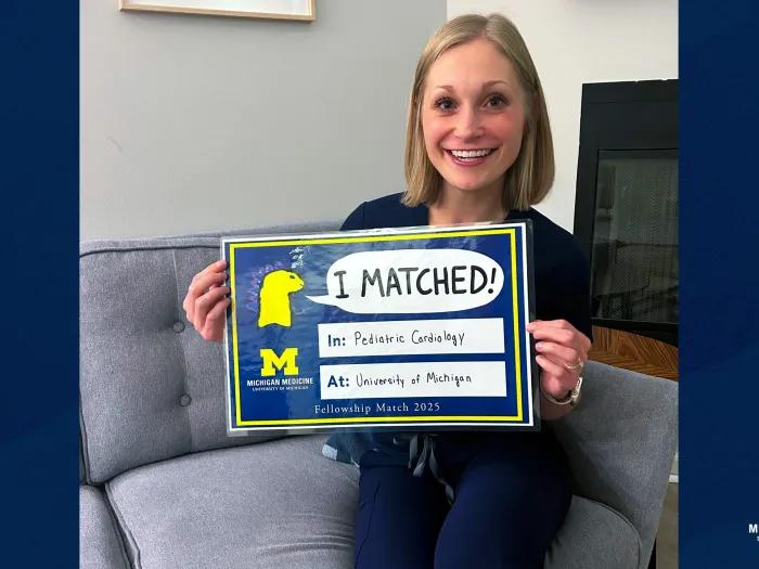
Fontan patient inspires as pediatric cardiology fellow
How lifesaving care after teen’s sudden cardiac arrest made motherhood possible years later