General Information About Childhood Vascular Tumors
Childhood vascular tumors can form anywhere in the body from abnormal blood vessel or lymph vessel cells.
These tumors may be benign (not cancer) or malignant (cancer). There are many types of vascular tumors. The most common type of childhood vascular tumor is infantile hemangioma, which is a benign tumor that usually goes away on its own.
Because malignant vascular tumors are rare in children, there is not a lot of information about what treatment works best.
Tests are used to diagnose childhood vascular tumors.
In addition to asking about your child's personal and family health history and doing a physical exam, your doctor may perform the following tests and procedures:
- Ultrasound exam: A procedure in which high-energy sound waves (ultrasound) are bounced off internal tissues or organs and make echoes. The echoes form a picture of body tissues called a sonogram. The picture can be printed to be looked at later.
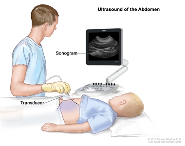
Abdominal ultrasound. An ultrasound transducer connected to a computer is pressed against the skin of the abdomen. The transducer bounces sound waves off internal organs and tissues to make echoes that form a sonogram (computer picture). - CT scan (CAT scan): A procedure that makes a series of detailed pictures of areas inside the body, taken from different angles. The pictures are made by a computer linked to an x-ray machine. A dye may be injected into a vein or swallowed to help the organs or tissues show up more clearly. This procedure is also called computed tomography, computerized tomography, or computerized axial tomography.
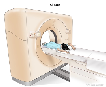
Computed tomography (CT) scan. The child lies on a table that slides through the CT scanner, which takes a series of detailed x-ray pictures of areas inside the body. - MRI (magnetic resonance imaging): A procedure that uses a magnet, radio waves, and a computer to make a series of detailed pictures of areas inside the body. This procedure is also called nuclear magnetic resonance imaging (NMRI).
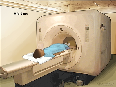
Magnetic resonance imaging (MRI) scan. The child lies on a table that slides into the MRI machine, which takes a series of detailed pictures of areas inside the body. The positioning of the child on the table depends on the part of the body being imaged. - X-ray: An x-ray of the bones or the chest. An x-ray is a type of energy beam that can go through the body onto film, making a picture of areas inside the body.
- Biopsy: The removal of cells or tissues so they can be viewed under a microscope by a pathologist to check for signs of cancer. A biopsy is not always needed to diagnose a vascular tumor.
Childhood vascular tumors may be classified into four groups.
Benign tumors
Benign tumors are not cancer. This summary has information about the following benign vascular tumors:
- Infantile hemangioma.
- Congenital hemangioma.
- Benign vascular tumors of the liver.
- Spindle cell hemangioma.
- Epithelioid hemangioma.
- Pyogenic granuloma (lobular capillary hemangioma).
- Angiofibroma.
- Juvenile nasopharyngeal angiofibroma.
Intermediate tumors that may spread locally
Some intermediate tumors are likely to spread to the area around the tumor (locally), but not to other parts of the body. This summary has information about the following tumors that spread locally:
- Kaposiform hemangioendothelioma and tufted angioma.
Intermediate tumors that may spread to other parts of the body
Rarely, intermediate tumors spread to other parts of the body (metastasize). This summary has information about the following vascular tumors that may metastasize:
- Pseudomyogenic (epithelioid sarcoma–like) hemangioendothelioma.
- Retiform hemangioendothelioma.
- Papillary intralymphatic angioendothelioma.
- Composite hemangioendothelioma.
- Kaposi sarcoma.
Malignant tumors
Malignant tumors are cancer. This summary has information about the following malignant vascular tumors:
- Epithelioid hemangioendothelioma.
- Angiosarcoma.
Treatment Option Overview
There are different types of treatment for childhood vascular tumors.
Different types of treatment are available for children with vascular tumors. Some treatments are standard (the currently used treatment), and some are being tested in clinical trials. A treatment clinical trial is a research study meant to help improve current treatments or obtain information on new treatments. When clinical trials show that a new treatment is better than the standard treatment, the new treatment may become the standard treatment.
Because vascular tumors in children are rare, taking part in a clinical trial should be considered. Some clinical trials are open only to patients who have not started treatment.
Children with childhood vascular tumors should have their treatment planned by a team of health care providers who are experts in treating cancer in children.
Treatment will be overseen by a pediatric oncologist, a doctor who specializes in treating children with cancer. The pediatric oncologist works with other pediatric health care providers who are experts in treating children with cancer and who specialize in certain areas of medicine. These may include the following specialists:
- Pediatric vascular anomaly specialist (expert in treating children with vascular tumors).
- Pediatric surgeon.
- Orthopedic surgeon.
- Radiation oncologist.
- Pediatric nurse specialist.
- Rehabilitation specialist.
- Psychologist.
- Social worker.
Twelve types of standard treatment are used:
Beta-blocker therapy
Beta-blockers are drugs that decrease blood pressure and heart rate. When used in patients with vascular tumors, beta-blockers may help shrink the tumors. Beta-blocker therapy may be given by vein (IV), by mouth, or placed on the skin (topical). The way the beta-blocker therapy is given depends on the type of vascular tumor and where the tumor first formed.
The beta-blocker propranolol is usually the first treatment for hemangiomas. Infants younger than 4 weeks, or who have an underlying condition, or who are treated with IV propranolol may need to have their treatment started in a hospital. Propranolol is also used to treat benign vascular tumor of liver and kaposiform hemangioendothelioma.
Other beta-blockers used to treat vascular tumors include atenolol, nadolol, and timolol.
Infantile hemangioma may also be treated with propranolol and steroid therapy or propranolol and topical beta-blocker therapy.
See the drug information summary on Propranolol Hydrochloride for more information.
Surgery
The following types of surgery may be used to remove many types of vascular tumors:
- Excision: Surgery to remove the entire tumor and some of the healthy tissue around it.
- Laser surgery: A surgical procedure that uses a laser beam (a narrow beam of intense light) as a knife to make bloodless cuts in tissue or to remove a skin lesion such as a tumor. Surgery with a pulsed dye laser may be used for some hemangiomas. This type of laser uses a beam of light that targets blood vessels in the skin. The light is changed into heat and the blood vessels are destroyed without damaging nearby skin.
- Curettage: A procedure in which abnormal tissue is removed using a small, spoon-shaped instrument with a sharp edge called a curette.
- Total hepatectomy and liver transplant: A surgical procedure to remove the entire liver followed by a transplant of a healthy liver from a donor.
The type of surgery used depends on the type of vascular tumor and where the tumor formed in the body.
For malignant tumors, after the doctor removes all the cancer that can be seen at the time of the surgery, some patients may be given chemotherapy or radiation therapy to kill any cancer cells that are left. Treatment given after the surgery, to lower the risk that the cancer will come back, is called adjuvant therapy.
Photocoagulation
Photocoagulation is the use of an intense beam of light, such as a laser, to seal off blood vessels or destroy tissue. It is used to treat pyogenic granuloma.
Cryotherapy
Cryotherapy is a treatment that uses an instrument to freeze and destroy abnormal tissue, such as abnormal blood vessels in pyogenic granuloma. This type of treatment is also called cryosurgery.
For more information, see Cryosurgery to Treat Cancer.
Embolization
Embolization is a procedure that uses particles, such as tiny gelatin sponges or beads, to block blood vessels in the liver. It may be used to treat some benign vascular tumors of the liver and kaposiform hemangioendothelioma.
Chemotherapy
Chemotherapy is a treatment that uses drugs to stop the growth of tumor cells, either by killing the cells or by stopping them from dividing. When chemotherapy is taken by mouth or injected into a vein or muscle, the drugs enter the bloodstream and can reach tumor cells throughout the body. Sometimes more than one anticancer drug is given. This is called combination chemotherapy.
Sclerotherapy
Sclerotherapy is a treatment used to destroy the blood vessel that leads to the tumor and the tumor. A liquid is injected into the blood vessel, causing it to scar and break down. Over time, the destroyed blood vessel is absorbed into normal tissue. The blood flows through nearby healthy veins instead. Sclerotherapy is used in the treatment of epithelioid hemangioma.
Radiation therapy
Radiation therapy is a treatment that uses high-energy x-rays or other types of radiation to kill tumor cells or keep them from growing. External radiation therapy uses a machine outside the body to send radiation toward the area of the body with the tumor. It is used to treat some vascular tumors.
Targeted therapy
Targeted therapy uses drugs or other substances to block the action of specific enzymes, proteins, or other molecules involved in the growth and spread of cancer cells. Different types of targeted therapy are being used or studied to treat childhood vascular tumors:
- Angiogenesis inhibitors: Angiogenesis inhibitors are drugs that stop cells from dividing and prevent the growth of new blood vessels that tumors need to grow. The targeted therapy drugs thalidomide, sorafenib, and pazopanib are angiogenesis inhibitors used to treat childhood vascular tumors.
- Mammalian target of rapamycin (mTOR) inhibitors: This treatment blocks a protein called mTOR, which may keep cancer cells from growing and prevent new blood vessels from forming. Sirolimus is an mTOR inhibitor used to treat childhood vascular tumors.
- Kinase inhibitors: Kinase inhibitors block signals needed for tumors to grow. Trametinib is being studied to treat epithelioid hemangioendothelioma.
Immunotherapy
Immunotherapy helps a person's immune system fight cancer.
The following types of immunotherapy are being used in the treatment of childhood vascular tumors:
Other drug therapy
Other drugs used to treat childhood vascular tumors or manage their effects include the following:
- Steroid therapy: Steroids are hormones made naturally in the body. They can also be made in a laboratory and used as drugs. Steroid drugs help shrink some vascular tumors. Corticosteroids, such as prednisone and methylprednisolone, are used to treat infantile hemangioma.
- Immunosuppressant therapy: These drugs decrease the body's immune responses. Immunosuppressant therapy has been used to help shrink vascular tumors. Topical tacrolimus is used to treat kaposiform hemangioendotheliomas and tufted angiomas.
- Thyroid hormone replacement therapy: Man-made hormones are used to treat a rare form of hypothyroidism caused by some vascular tumors, such as liver hemangiomas.
Observation
Observation is closely monitoring a patient's condition without giving any treatment until signs or symptoms appear or change.
New types of treatment are being tested in clinical trials.
Information about clinical trials is available from the NCI website.
Treatment for childhood vascular tumors may cause side effects.
For information about side effects that begin during treatment for cancer, see our Side Effects page.
Some treatments, such as chemotherapy and radiation therapy, cause side effects that continue or appear months or years after treatment has ended. These are called late effects. Late effects of treatment may include the following:
- Physical problems.
- Changes in mood, feelings, thinking, learning, or memory.
- Second cancers (new types of cancer).
Some late effects may be treated or controlled. It is important to talk with your child's doctors about the possible late effects caused by some treatments. For more information, see Late Effects of Treatment for Childhood Cancer.
Patients may want to think about taking part in a clinical trial.
For some patients, taking part in a clinical trial may be the best treatment choice. Clinical trials are part of the research process. Clinical trials are done to find out if new treatments are safe and effective or better than the standard treatment.
Many of today's standard treatments are based on earlier clinical trials. Patients who take part in a clinical trial may receive the standard treatment or be among the first to receive a new treatment.
Patients who take part in clinical trials also help improve the way disease will be treated in the future. Even when clinical trials do not lead to effective new treatments, they often answer important questions and help move research forward.
Patients can enter clinical trials before, during, or after starting their tumor treatment.
Some clinical trials only include patients who have not yet received treatment. Other trials test treatments for patients whose tumors have not gotten better. There are also clinical trials that test new ways to stop tumors from recurring (coming back) or reduce the side effects of treatment.
Clinical trials are taking place in many parts of the country. Information about clinical trials supported by NCI can be found on NCI's clinical trials search webpage. Clinical trials supported by other organizations can be found on the ClinicalTrials.gov website.
Follow-up tests may be needed.
Some of the tests that were done to diagnose the vascular tumor may be repeated. Some tests will be repeated to see how well the treatment is working. Decisions about whether to continue, change, or stop treatment may be based on the results of these tests.
Some of the tests will continue to be done from time to time after treatment has ended. The results of these tests can show if your child's condition has changed or if the tumor has recurred (come back). These tests are sometimes called follow-up tests or check-ups.
Benign Tumors
For information about the treatments listed below, see the Treatment Option Overview section.
Infantile Hemangioma
Infantile hemangiomas are the most common type of benign vascular tumor in children. Infantile hemangiomas form when immature cells that are meant to form blood vessels form a tumor instead. An infantile hemangioma may also be called a "strawberry mark."
These tumors are not usually seen at birth but appear when the infant is 3 to 6 weeks old. Most hemangiomas get bigger for about 5 months and then stop growing. The hemangiomas slowly fade away over the next several years, but a red mark or loose or wrinkled skin may remain. It is rare for an infantile hemangioma to come back after it has faded away.
Infantile hemangiomas may be on the skin, in the tissue below the skin, and/or in an organ. They are usually on the head and neck but can be anywhere on or in the body. Hemangiomas may appear as a single lesion, one or more lesions spread over a larger area of the body, or multiple lesions in more than one part of the body. Lesions that are spread over a larger area of the body or multiple lesions are more likely to cause problems.
Infantile hemangioma with minimal or arrested growth (IH-MAG) is a certain type of infantile hemangioma that is seen at birth and does not tend to get bigger. The lesion appears as light and dark areas of redness in the skin. The lesions are usually on the lower body but can be on the head and neck. Hemangiomas of this type go away over time without treatment.
Risk Factors
A risk factor is anything that increases the chance of getting a disease. Not every child with a risk factor will develop infantile hemangiomas, and they can develop in some children who don't have a known risk factor. Talk with your child's doctor if you think your child may be at risk of an infantile hemangioma.
Infantile hemangiomas are more common in the following:
- Girls.
- White patients.
- Premature babies.
- Twins, triplets, or other multiple births.
- Babies of mothers who are older at the time of the pregnancy, have pre-eclampsia (high blood pressure during pregnancy), or who have problems with the placenta during pregnancy.
Other risk factors for infantile hemangiomas include the following:
- Having a family history of infantile hemangioma, usually in a mother, father, sister, or brother.
- Having certain syndromes.
- PHACE syndrome: A syndrome in which the hemangioma spreads across a large area of the head or face and sometimes the neck, chest, or arm. Other health problems that affect the large blood vessels, heart, eyes, and/or brain may also occur.
- LUMBAR/PELVIS/SACRAL syndrome: A syndrome in which the hemangioma spreads across a large area of the lower back. Other health problems that affect the urinary system, genitals, rectum, anus, brain, spinal cord, and nerve function may also occur.
Having more than one hemangioma or a hemangioma in the airway or eyes increases the risk of having other health problems.
- Multiple hemangiomas: Having more than five hemangiomas on the skin is a sign that there may be hemangiomas in an organ. The liver is affected most often. Heart, muscle, and thyroid gland problems can also occur.
- Airway hemangiomas: Hemangiomas in the airway usually occur along with a large, beard-shaped area of hemangioma on the face (from the ears, around the mouth, lower chin, and front of neck). It is important that airway hemangiomas are treated before the child has trouble breathing.
- Ophthalmic hemangiomas: Hemangiomas that involve the eye may cause vision problems or blindness. Infantile hemangiomas can occur in the conjunctiva (a membrane that lines the inner surface of the eyelid and covers the front part of the eye). These hemangiomas may be linked with other abnormal conditions of the eye. It is important that children with an ophthalmic hemangioma are examined by an ophthalmologist.
Signs and Symptoms
Infantile hemangiomas may cause any of the following signs and symptoms. Check with your child's doctor if your child has any of the following:
- Skin lesions: An area of spidery veins or lightened or discolored skin may appear before the hemangioma does. Hemangiomas occur as firm, warm, bright red to crimson lesions on the skin or may look like a bruise. Lesions that form ulcers are also painful. Later, as the hemangiomas go away, they become softer and begin fading in the center before flattening and losing color.
- Lesions below the skin: Lesions that grow under the skin in the fat may appear blue or purple. If the lesions are deep enough under the skin surface, they may not be seen.
- Lesions in an organ: There may be no signs that hemangiomas have formed on an organ.
Although most infantile hemangiomas are nothing to worry about, if your child develops any lumps or red or blue marks on the skin check with your child's doctor. He or she can refer the child to a specialist if needed.
Diagnostic Tests
A physical exam and history are usually all that are needed to diagnose infantile hemangiomas. If there is something about the tumor that looks unusual, a biopsy may be done. If the hemangioma is deeper inside the body with no change to the skin, or the lesions are spread across a large area of the body, an ultrasound may be done. Infants with five or more hemangiomas on the skin should have an ultrasound of the liver done to check for a liver hemangioma. See the General Information section for a description of these tests and procedures.
If the hemangiomas are part of a syndrome, more tests may be done, such as an echocardiogram, MRI, magnetic resonance angiogram, and eye exam.
Treatment
Most hemangiomas fade and shrink without treatment. If the hemangioma is large or causing other health problems, treatment may include the following:
- Propranolol or other beta-blocker therapy.
- Steroid therapy, before beta-blocker therapy is begun or when beta-blockers cannot be used.
- Laser surgery, including pulsed dye laser surgery, may be used for hemangiomas that have ulcers or have not completely gone away.
- Surgery for hemangiomas that have ulcers, cause vision problems, or have not completely gone away. Surgery may also be used for lesions on the face that do not respond to other treatment.
- Topical beta-blocker therapy for hemangiomas that are in one area of the skin.
- Combined therapy, such as propranolol and steroid therapy or propranolol and topical beta-blocker therapy.
- A clinical trial of propranolol.
Congenital Hemangioma
Congenital hemangioma is a benign vascular tumor that begins forming before birth and is fully formed when the baby is born. They're usually on the skin but can be in another organ. A congenital hemangioma may occur as a rash of purple spots and the skin around the spot may be lighter.
There are three types of congenital hemangiomas:
- Rapidly involuting congenital hemangioma: These tumors go away on their own 12 to 15 months after birth. They can form ulcers, bleed, and cause temporary heart and blood clotting problems. The skin may look a little different even after the hemangiomas go away.
- Partial involuting congenital hemangioma: These tumors do not go away completely.
- Non-involuting congenital hemangioma: These tumors never go away on their own.
Diagnostic Tests
See the General Information section for a description of tests and procedures, such as ultrasound, used to diagnose congenital hemangioma.
Treatment
Treatment of rapidly involuting congenital hemangioma and partial involuting congenital hemangioma may include the following:
Treatment of non-involuting congenital hemangioma may include the following:
- Surgery to remove the tumor, depending on where it is and whether it is causing symptoms.
Benign Vascular Tumors of the Liver
Benign vascular tumors of the liver may be focal vascular lesions (a single lesion in one area of the liver), multiple liver lesions (more than one lesion in one area of the liver), or diffuse liver lesions (more than one lesion in more than one area of the liver).
The liver has many functions, including filtering blood and making proteins needed for blood clotting. Sometimes, blood that normally flows through the liver is blocked or slowed by the tumor. This sends blood directly to the heart without going through the liver and is called a liver shunt. This can cause heart failure and problems with blood clotting.
Focal Vascular Lesions
Focal vascular lesions are usually rapidly involuting congenital hemangiomas or non-involuting congenital hemangiomas.
Diagnostic Tests
See the General Information section for a description of tests and procedures used to diagnose focal vascular lesions of the liver.
Treatment
Treatment of focal vascular lesions of the liver depends on whether symptoms occur and may include the following:
- Observation.
- Embolization of the liver to treat symptoms.
- Surgery, for lesions that do not respond to other treatments.
Multiple and Diffuse Liver Lesions
Multiple and diffuse lesions of the liver are usually infantile hemangiomas. Diffuse lesions of the liver can cause serious effects, including problems with the thyroid gland and heart. The liver can enlarge, press on other organs, and cause more symptoms.
Diagnostic Tests
See the General Information section for a description of tests and procedures used to diagnose multiple or diffuse benign vascular lesions.
Treatment
Treatment of multiple liver lesions may include the following:
- Observation for lesions that do not cause symptoms.
- Beta-blocker therapy (propranolol) for lesions that begin to grow.
Treatment of diffuse liver lesions may include the following:
- Beta-blocker therapy (propranolol).
- Thyroid hormone replacement therapy.
- Chemotherapy.
- Steroid therapy.
- Total hepatectomy and liver transplant, when the lesions do not respond to drug therapy. This is done only when the lesions have spread widely in the liver and more than one organ has failed.
If a vascular lesion of the liver does not respond to standard treatments, a biopsy may be done to see if the tumor has become cancer.
Spindle Cell Hemangioma
Spindle cell hemangiomas contain cells called spindle cells. Under a microscope, spindle cells look long and slender.
Risk Factors
A risk factor is anything that increases the chance of getting a disease. Not every child with a risk factor will develop spindle cell hemangiomas, and they can develop in some children who don't have a known risk factor. Talk with your child's doctor if you think your child may be at risk. Spindle cell hemangiomas are likely to occur in children with the following syndromes:
- Maffucci syndrome, which affects cartilage and skin.
- Klippel-Trenaunay syndrome, which affects blood vessels, soft tissues, and bones.
Signs
Spindle cell hemangiomas appear on or under the skin. They are painful red-brown or bluish lesions that usually appear on the arms or legs. They can begin as one lesion and develop into more lesions over years.
Diagnostic Tests
See the General Information section for a description of tests and procedures used to diagnose spindle cell hemangioma.
Treatment
There is no standard treatment for spindle cell hemangiomas. Treatment may include the following:
- Surgery to remove the tumor.
Spindle cell hemangiomas may come back after surgery.
Epithelioid Hemangioma
Epithelioid hemangiomas usually form on or in the skin, especially the head, but can occur in other areas, such as bone.
Signs and Symptoms
Epithelioid hemangiomas are sometimes caused by injury. On the skin, they may appear as firm pink to red bumps and may be itchy. Epithelioid hemangioma of the bone may cause swelling, pain, and weakened bone in the affected area.
Diagnostic Tests
See the General Information section for a description of tests and procedures used to diagnose epithelioid hemangioma.
Treatment
There is no standard treatment for epithelioid hemangiomas. Treatment may include the following:
- Surgery (curettage or resection).
- Sclerotherapy.
- Radiation therapy in rare cases.
Epithelioid hemangiomas often come back after treatment.
Pyogenic Granuloma
Pyogenic granuloma is also called lobular capillary hemangioma. It is most common in older children and young adults but may occur at any age.
The lesions are sometimes caused by injury or from the use of certain medicines, including birth control pills and retinoids. They may also form for no known reason inside capillaries (the smallest blood vessels), arteries, veins, or other places on the body. Some lesions may be associated with capillary malformations. Usually there is only one lesion, but sometimes multiple lesions occur in the same area or the lesions may spread to other areas of the body.
Signs
Pyogenic granulomas are raised, bright red lesions that may be small or large and smooth or bumpy. They grow quickly over weeks to months and may bleed a lot. These lesions are usually on the surface of the skin, but may form in the tissues below the skin and look like other vascular lesions.
Diagnostic Tests
See the General Information section for a description of tests and procedures used to diagnose pyogenic granuloma.
Treatment
Some pyogenic granulomas go away without treatment. Other pyogenic granulomas need treatment that may include the following:
- Surgery (excision or curettage) to remove the lesion.
- Photocoagulation.
- Cryotherapy.
- Topical beta-blocker therapy (propranolol or timolol).
Pyogenic granulomas often come back after treatment.
Angiofibroma
Angiofibromas are rare. They are benign skin lesions that usually occur with a condition called tuberous sclerosis (an inherited disorder that causes skin lesions, seizures, and mental disabilities).
Signs
Angiofibromas appear as red bumps on the face.
Diagnostic Tests
See the General Information section for a description of tests and procedures used to diagnose angiofibroma.
Treatment
Treatment of angiofibromas may include the following:
- Surgery to remove the tumor.
- Laser therapy.
- Targeted therapy (sirolimus).
Juvenile Nasopharyngeal Angiofibroma
Juvenile nasopharyngeal angiofibromas are benign tumors but they can grow into nearby tissue. They begin in the nasal cavity and may spread to the nasopharynx, the paranasal sinuses, the bone around the eyes, and sometimes to the brain.
Diagnostic Tests
See the General Information section for a description of tests and procedures used to diagnose juvenile nasopharyngeal angiofibroma.
Treatment
Treatment of juvenile nasopharyngeal angiofibromas may include the following:
- Surgery to remove the tumor.
- Radiation therapy.
- Chemotherapy.
- Immunotherapy (interferon).
- Targeted therapy (sirolimus).
Intermediate Tumors that May Spread Locally
For information about the treatments listed below, see the Treatment Option Overview section.
Kaposiform Hemangioendothelioma and Tufted Angioma
Kaposiform hemangioendotheliomas and tufted angiomas are blood vessel tumors that occur in infants or young children. These tumors can cause Kasabach-Merritt phenomenon, a condition in which the blood is not able to clot and serious bleeding may occur. In Kasabach-Merritt phenomenon, the tumor traps and destroys platelets (blood-clotting cells). Then there aren't enough platelets in the blood when needed to stop bleeding. This type of vascular tumor is not related to Kaposi sarcoma.
Signs and Symptoms
Kaposiform hemangioendotheliomas and tufted angiomas usually occur on the skin of the arms and legs, but may also form in deeper tissues, such as muscle or bone, or in the chest, abdomen, head, or neck.
Signs and symptoms may include the following:
- Firm, painful areas of skin that look bruised.
- Purple or brownish-red areas of skin.
- Pain with no visible lump.
- Easy bruising.
- Bleeding more than the usual amount from mucous membranes, wounds, and other tissues.
Patients who have kaposiform hemangioendothelioma and tufted angioma may have anemia (weakness, feeling tired, or looking pale).
Diagnostic Tests
See the General Information section for a description of tests and procedures used to diagnose kaposiform hemangioendothelioma and tufted angioma.
If a physical exam and MRI clearly show the tumor is a kaposiform hemangioendothelioma or a tufted angioma, a biopsy may not be needed. A biopsy is not always done because serious bleeding can occur.
Treatment
Treatment of kaposiform hemangioendotheliomas and tufted angiomas depends on the child's symptoms. Infection, delay in treatment, and surgery can cause bleeding that is life-threatening. Kaposiform hemangioendotheliomas and tufted angiomas are best treated by a vascular anomaly specialist.
Treatment for uncomplicated kaposiform hemangioendotheliomas and tufted angiomas may include the following:
- Observation.
- Surgery to remove the tumor.
- Laser surgery.
- Topical therapy (steroids or tacrolimus).
- Beta-blocker therapy (propranolol).
- Targeted therapy (sirolimus).
Treatment for complicated kaposiform hemangioendotheliomas and tufted angiomas may include the following:
- Chemotherapy, with or without steroid therapy.
- Targeted therapy (sirolimus), with or without steroid therapy.
- Surgery, with or without embolization.
Even with treatment, these tumors do not fully go away and can come back. Pain and inflammation may get worse with age, often around the time of puberty. Long-term effects include chronic pain, heart failure, bone problems, and lymphedema (the build up of lymph fluid in tissues).
Intermediate Tumors that May Spread to Other Parts of the Body
For information about the treatments listed below, see the Treatment Option Overview section.
Pseudomyogenic (Epithelioid Sarcoma–Like) Hemangioendothelioma
Pseudomyogenic hemangioendothelioma can occur in children, but is most common in men aged 20 to 50 years. These tumors are rare, and usually occur on or under the skin, or in bone. They may spread to nearby tissue but usually do not spread to other parts of the body. In most cases, there are multiple tumors.
Signs and Symptoms
Pseudomyogenic hemangioendotheliomas may appear as a lump in soft tissue or may cause pain in the affected area.
Diagnostic Tests
See the General Information section for a description of tests and procedures used to diagnose pseudomyogenic hemangioendothelioma.
Treatment
Treatment of pseudomyogenic hemangioendotheliomas may include the following:
- Surgery to remove the tumor when possible. Amputation may be needed when there are multiple tumors in the bone.
- Chemotherapy.
- Targeted therapy (mTOR inhibitors).
Because pseudomyogenic hemangioendothelioma is so rare in children, treatment options are based on clinical trials in adults.
Retiform Hemangioendothelioma
Retiform hemangioendotheliomas are slow growing, flat tumors that occur in young adults and sometimes children. These tumors usually occur on or under the skin of the arms, legs, and trunk. These tumors usually do not spread to other parts of the body.
Diagnostic Tests
See the General Information section for a description of tests and procedures used to diagnose retiform hemangioendothelioma.
Treatment
Treatment of retiform hemangioendotheliomas may include the following:
- Surgery to remove the tumor. Follow-up will include monitoring to see if the tumor comes back.
- Radiation therapy and chemotherapy when surgery cannot be done or when the tumor has come back.
Retiform hemangioendothelioma may come back after treatment.
Papillary Intralymphatic Angioendothelioma
Papillary intralymphatic angioendotheliomas are also called Dabska tumors. These tumors form in or under the skin anywhere on the body. Lymph nodes are sometimes affected.
Signs
Papillary intralymphatic angioendotheliomas may appear as firm, raised, purplish bumps, which may be small or large.
Diagnostic Tests
See the General Information section for a description of tests and procedures used to diagnose papillary intralymphatic angioendothelioma.
Treatment
Treatment of papillary intralymphatic angioendotheliomas may include the following:
- Surgery to remove the tumor.
Composite Hemangioendothelioma
Composite hemangioendotheliomas have features of both benign and malignant vascular tumors. These tumors usually occur on or under the skin on the arms or legs. They may also occur on the head, neck, or chest. Composite hemangioendotheliomas are not likely to metastasize (spread) but they may come back in the same place. When the tumors metastasize, they usually spread to nearby lymph nodes.
Diagnostic Tests
See the General Information section for a description of tests and procedures used to diagnose composite hemangioendothelioma and find out whether the tumor has spread.
Treatment
Treatment of composite hemangioendotheliomas may include the following:
- Surgery to remove the tumor.
- Radiation therapy and chemotherapy for tumors that have spread.
Kaposi Sarcoma
Kaposi sarcoma is a cancer that causes lesions to grow in the skin; the mucous membranes lining the mouth, nose, and throat; lymph nodes; or other organs. It is caused by the Kaposi sarcoma herpes virus (KSHV). In the United States, it occurs most often in children who have a weak immune system caused by rare immune system disorders, HIV infection, or drugs used in organ transplants.
Signs
Signs in children may include the following:
- Lesions in the skin, mouth, or throat. Skin lesions are red, purple, or brown and change from flat, to raised, to scaly areas called plaques, to nodules.
- Swollen lymph nodes.
Diagnostic Tests
See the General Information section for a description of tests and procedures used to diagnose Kaposi sarcoma.
Treatment
Treatment of Kaposi sarcoma may include the following:
- Chemotherapy.
- Immunotherapy (interferon).
- Radiation therapy.
- Antiretroviral therapy.
Because Kaposi sarcoma is so rare in children, some treatment options are based on clinical trials in adults. For more information about Kaposi sarcoma in adults, see Kaposi Sarcoma Treatment.
Malignant Tumors
For information about the treatments listed below, see the Treatment Option Overview section.
Epithelioid Hemangioendothelioma
Epithelioid hemangioendotheliomas can occur in children, but are most common in adults aged 30 to 50 years. They usually occur in the liver, lung, or in bone. They may be fast growing or slow growing. In about a third of cases, the tumor spreads to other parts of the body very quickly.
Signs and Symptoms
Signs and symptoms depend on where the tumor is in the body. Check with your child's doctor if your child has any of the following:
- On the skin, the tumors can be raised and rounded or flat, red-brown patches that feel warm.
- In the lung, there may be no early symptoms. Signs and symptoms that may occur include:
- Chest pain.
- Spitting up blood.
- Anemia (weakness, feeling tired, or looking pale).
- Trouble breathing (from scarred lung tissue).
- In bone, the tumors can cause breaks.
Tumors that occur in the liver or soft tissue may also cause signs and symptoms.
Diagnostic Tests
Epithelioid hemangioendotheliomas in the liver are found with CT scans and MRI scans. See the General Information section for a description of these tests and procedures used to diagnose epithelioid hemangioendothelioma and find out whether the tumor has spread. X-rays may also be done.
Treatment
Treatment of slow-growing epithelioid hemangioendotheliomas includes the following:
Treatment of fast-growing epithelioid hemangioendotheliomas may include the following:
- Surgery to remove the tumor when possible.
- Immunotherapy (interferon) and targeted therapy (thalidomide, sorafenib, pazopanib, sirolimus) for tumors that are likely to spread.
- Chemotherapy.
- Radiation therapy.
- Total hepatectomy and liver transplant when the tumor is in the liver.
Angiosarcoma
Angiosarcomas are fast-growing tumors that form in blood vessels or lymph vessels in any part of the body, usually in soft tissue. Most angiosarcomas are in or near the skin. Those in deeper soft tissue can form in the liver, spleen, and lung.
These tumors are very rare in children. Children sometimes have more than one tumor in the skin and/or liver.
Risk Factors
A risk factor is anything that increases the chance of getting a disease. Not every child with a risk factor will develop angiosarcoma, and it can develop in some children who don't have a known risk factor. Talk with your child's doctor if you think your child may be at risk. Risk factors for angiosarcomas include the following:
- Being exposed to radiation.
- Chronic (long-term) lymphedema, a condition in which extra lymph fluid builds up in tissues and causes swelling.
- Having a benign vascular tumor. A benign tumor, such as a hemangioma, may become an angiosarcoma but this is rare.
Signs
Signs of angiosarcoma depend on where the tumor is and may include the following:
- Red patches on the skin that bleed easily.
- Purple tumors.
Diagnostic Tests
See the General Information section for a description of tests and procedures used to diagnose angiosarcoma and find out whether the tumor has spread.
Treatment
Treatment of angiosarcoma may include the following:
- Surgery to completely remove the tumor.
- A combination of surgery, chemotherapy, and radiation therapy for angiosarcomas that have spread.
- Targeted therapy (bevacizumab) and chemotherapy for angiosarcomas that began as infantile hemangiomas.
- A clinical trial of immunotherapy (nivolumab and ipilimumab).
To Learn More About Childhood Vascular Tumors
For more information about childhood vascular tumors, see the following:
- Soft Tissue Sarcoma Home Page
- Computed Tomography (CT) Scans and Cancer
- Targeted Therapy to Treat Cancer
- Immunotherapy to Treat Cancer
- MyPART - My Pediatric and Adult Rare Tumor Network
For more childhood cancer information and other general cancer resources, see the following:
- About Cancer
- Childhood Cancers
- CureSearch for Children's Cancer
- Late Effects of Treatment for Childhood Cancer
- Adolescents and Young Adults with Cancer
- Children with Cancer: A Guide for Parents
- Cancer in Children and Adolescents
- Staging
- Coping with Cancer
- Questions to Ask Your Doctor about Cancer
- For Survivors and Caregivers
About This PDQ Summary
About PDQ
Physician Data Query (PDQ) is the National Cancer Institute's (NCI's) comprehensive cancer information database. The PDQ database contains summaries of the latest published information on cancer prevention, detection, genetics, treatment, supportive care, and complementary and alternative medicine. Most summaries come in two versions. The health professional versions have detailed information written in technical language. The patient versions are written in easy-to-understand, nontechnical language. Both versions have cancer information that is accurate and up to date and most versions are also available in Spanish.
PDQ is a service of the NCI. The NCI is part of the National Institutes of Health (NIH). NIH is the federal government's center of biomedical research. The PDQ summaries are based on an independent review of the medical literature. They are not policy statements of the NCI or the NIH.
Purpose of This Summary
This PDQ cancer information summary has current information about the treatment of childhood vascular tumors. It is meant to inform and help patients, families, and caregivers. It does not give formal guidelines or recommendations for making decisions about health care.
Reviewers and Updates
Editorial Boards write the PDQ cancer information summaries and keep them up to date. These Boards are made up of experts in cancer treatment and other specialties related to cancer. The summaries are reviewed regularly and changes are made when there is new information. The date on each summary ("Updated") is the date of the most recent change.
The information in this patient summary was taken from the health professional version, which is reviewed regularly and updated as needed, by the PDQ Pediatric Treatment Editorial Board.
Clinical Trial Information
A clinical trial is a study to answer a scientific question, such as whether one treatment is better than another. Trials are based on past studies and what has been learned in the laboratory. Each trial answers certain scientific questions in order to find new and better ways to help cancer patients. During treatment clinical trials, information is collected about the effects of a new treatment and how well it works. If a clinical trial shows that a new treatment is better than one currently being used, the new treatment may become "standard." Patients may want to think about taking part in a clinical trial. Some clinical trials are open only to patients who have not started treatment.
Clinical trials can be found online at NCI's website. For more information, call the Cancer Information Service (CIS), NCI's contact center, at 1-800-4-CANCER (1-800-422-6237).
Permission to Use This Summary
PDQ is a registered trademark. The content of PDQ documents can be used freely as text. It cannot be identified as an NCI PDQ cancer information summary unless the whole summary is shown and it is updated regularly. However, a user would be allowed to write a sentence such as "NCI's PDQ cancer information summary about breast cancer prevention states the risks in the following way: [include excerpt from the summary]."
The best way to cite this PDQ summary is:
PDQ® Pediatric Treatment Editorial Board. PDQ Childhood Vascular Tumors Treatment. Bethesda, MD: National Cancer Institute. Updated <MM/DD/YYYY>. Available at: https://www.cancer.gov/types/soft-tissue-sarcoma/patient/child-vascular-tumors-treatment-pdq. Accessed <MM/DD/YYYY>. [PMID: 27253005]
Images in this summary are used with permission of the author(s), artist, and/or publisher for use in the PDQ summaries only. If you want to use an image from a PDQ summary and you are not using the whole summary, you must get permission from the owner. It cannot be given by the National Cancer Institute. Information about using the images in this summary, along with many other images related to cancer can be found in Visuals Online. Visuals Online is a collection of more than 3,000 scientific images.
Disclaimer
The information in these summaries should not be used to make decisions about insurance reimbursement. More information on insurance coverage is available on Cancer.gov on the Managing Cancer Care page.
Contact Us
More information about contacting us or receiving help with the Cancer.gov website can be found on our Contact Us for Help page. Questions can also be submitted to Cancer.gov through the website's E-mail Us.
Last Revised: 2023-07-27
If you want to know more about cancer and how it is treated, or if you wish to know about clinical trials for your type of cancer, you can call the NCI's Cancer Information Service at 1-800-422-6237, toll free. A trained information specialist can talk with you and answer your questions.