Bone Density
Test Overview
A bone density test is a kind of X-ray test. It measures the density of minerals (such as calcium) in your bones. This information helps your doctor estimate the strength of your bones.
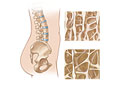
We all lose some bone mass as we age. Bones naturally become thinner as you grow older. This is because existing bone tissue is broken down faster than new bone is made. As this occurs, our bones lose calcium and other minerals. They also become lighter and less dense. This makes the bones weaker and makes them more likely to break (fracture).
With further bone loss, low bone density (sometimes called osteopenia) can lead to osteoporosis. So the thicker your bones are, the longer it takes to get osteoporosis. Although osteoporosis can occur in men, it is most common in women older than age 65.
If your bone density is lower than normal, you can increase it and your strength. You can do things like exercising, lifting weights or using weight machines. You can also make sure to get enough calcium and vitamin D. And you may need to take certain medicines.
There are several different ways to measure bone density.
- Dual-energy X-ray absorptiometry (DXA). This is the most accurate way to measure bone density. It uses two different X-ray beams to estimate bone density in your spine and hip. Strong, dense bones allow less of the X-ray beam to pass through them. The amounts of each X-ray beam that are blocked by bone and soft tissue are compared to each other. DXA can measure as little as 2% of bone loss per year. It is fast and uses very low doses of radiation. Single-energy X-ray absorptiometry (SXA) may be used to measure bone density in the heel and forearm. But SXA is not used as often as DXA.
- Peripheral dual-energy X-ray absorptiometry (P-DXA). P-DXA is a type of DXA test. It measures the density of bones in the arms or legs, such as the wrist. It can't measure the density of the bones most likely to break, such as the hip and spine. P-DXA machines are portable units that can be used in a doctor's office. P-DXA also uses very low doses of radiation. The results are ready faster than standard DXA measurements. P-DXA is not as useful as DXA for finding out how well medicine used to treat osteoporosis is working.
- Dual photon absorptiometry (DPA). This test uses a radioactive substance to measure bone density. It can measure bone density in your hip and spine. DPA also uses very low doses of radiation. But the scan takes longer than the other methods.
Ultrasound is a screening test that is sometimes offered at events such as health fairs. If results from an ultrasound test find low bone density, DXA is advised to confirm the results. Ultrasound uses sound waves to measure bone density, usually in your heel. It is quick and painless. And it does not use potentially harmful radiation like X-rays. One downside of ultrasound is that it can't measure the density of the bones in the hip and spine. These are the bones most likely to fracture from osteoporosis. Ultrasound is not used to keep track of how well medicine for osteoporosis is working.
Before you are screened for osteoporosis, you may want to think about what you will do if the tests show that you have a high chance of getting osteoporosis.
Health Tools
Health Tools help you make wise health decisions or take action to improve your health.
Why It Is Done
Doctors may recommend bone density tests for:footnote 1, footnote 2
- All women who are age 65 or older.
- Women younger than 65 who are at higher risk for broken bones caused by osteoporosis.
- Certain men at higher risk for osteoporosis.
- Follow-up of how well treatment for osteoporosis is working for men and women being treated for 2 years or longer.
How To Prepare
Avoid wearing clothes with metal buttons or buckles. You also may want to remove any jewelry that might cause a problem with the scan. For instance, don't wear a bracelet if you are having the scan done on your wrist.
How It Is Done
In most cases, a bone density scan is done in a radiology department or clinic by a technologist. Peripheral dual-energy X-ray absorptiometry (P-DXA) machines are portable units that can be used in a doctor's office.
You will need to lie on your back on a padded table. You probably can leave your clothes on. You may need to lie with your legs straight. Or you may lie with your lower legs resting on a platform built into the table.
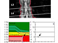
The machine will scan your bones and measure the amount of radiation they absorb. The DXA scan, which scans the hip and lower spine, takes about 20 minutes. Other kinds of scans may take 30 to 45 minutes.
Portable machines (P-DXA) can measure bone density in the wrist or forearm.
Testing at least two different bones each time is the most reliable way of measuring bone density. The hip and spine bones are preferred. It is best to test the same bones each time. The same type of scan and bone density equipment should also be used.
How It Feels
A bone density test does not cause pain. But if you have back pain, it may bother you to lie still on a table during the scan.
Risks
During a bone density scan, you are exposed to a very low dose of radiation. A bone density scan is not advised for pregnant women because it exposes the unborn baby to radiation.
Results
A bone density test is a kind of X-ray test. It measures the density of minerals (such as calcium) in your bones. Results are usually ready in 2 to 3 days.
Results of bone density tests can be reported in several ways.
T-score
Your T-score is your bone density compared to the average score of a healthy 30-year-old. (This is called the young adult reference range). It is expressed as a standard deviation (SD).
- A negative (–) value means that you have thinner bones (lower bone density) than an average 30-year-old. The more negative the number is, the less bone density you have compared with an average 30-year-old.
- A positive (+) value means that your bones are thicker and stronger than an average 30-year-old's.
The following table contains the World Health Organization's definitions of osteoporosis based on bone density T-scores.
T-score | |
Normal: | Less than 1 standard deviation (SD) below the young adult reference range (more than –1) |
Low bone density (osteopenia): | 1 to 2.5 SDs below the young adult reference range (–1 to –2.5) |
Osteoporosis: | More than 2.5 SDs below the young adult reference range (–2.5 or less) |
If your bone density test result is low:
- You may have osteoporosis. Doctors usually use the lowest T-score to diagnose osteoporosis. For example, if your T-score at your spine is –3 and your T-score at your hip is –2, the spine T-score would be used to diagnosis osteoporosis.
- Your chance of breaking a bone is higher than average. The more negative your T-score, the greater your chances of breaking a bone during a fall or from a minor injury. Every change of 1 SD means you have double the chance of a break at that site. For example, if you have a T-score of –1, your chances of having a broken bone are 2 times greater than if your T-score was 0.
Low bone density values may be caused by other problems, such as:
- Taking certain medicines.
- Cancer, such as multiple myeloma.
- Cushing's syndrome, hyperthyroidism, and hyperparathyroidism.
- Diseases of the spine, such as ankylosing spondylitis.
- Early menopause.
- Low levels of vitamin D.
- Heavy alcohol use.
Z-score
Your bone density value may also be compared to other people of your age, sex, and race. This is called your Z-score. It is given in standard deviations (SD) from the average value for your age group.
- A negative (–) value means that your bones are thinner (lower bone density) and weaker than most people in your age group. The more negative the number is, the less bone density you have compared with others in your age group.
- A positive (+) value means that your bones are thicker and stronger than most people in your age group.
What Affects the Test
You may not be able to have the test, or the results may not be helpful, if:
- You aren't able to be put in the correct position during the test.
- You had a broken bone in the past. This can cause falsely high bone density results.
- You have arthritis of your spine. The changes caused by arthritis in the spine may not make the spine the best place to measure for osteoporosis.
- You have metal implants from hip replacement surgery or hip fracture.
- You had an X-ray test that uses barium within 10 days of the bone density test.
References
Citations
- U.S. Preventive Services Task Force, et al. (2018). Screening for osteoporosis to prevent fractures: U.S. Preventive Services Task Force recommendation statement. JAMA, 319(24): 2521–2531. DOI: 10.1001/jama.2018.7498. Accessed October 29, 2018.
- National Osteoporosis Foundation (2014). Clinician's guide to prevention and treatment of osteoporosis. National Osteoporosis Foundation. http://nof.org/hcp/clinicians-guide. Accessed October 22, 2014.
- Fischbach FT, Dunning MB III, eds. (2009). Manual of Laboratory and Diagnostic Tests, 8th ed. Philadelphia: Lippincott Williams and Wilkins.
Credits
Current as of: September 25, 2023
Author: Healthwise Staff
Clinical Review Board
All Healthwise education is reviewed by a team that includes physicians, nurses, advanced practitioners, registered dieticians, and other healthcare professionals.
Current as of: September 25, 2023
Author: Healthwise Staff
Clinical Review Board
All Healthwise education is reviewed by a team that includes physicians, nurses, advanced practitioners, registered dieticians, and other healthcare professionals.
This information does not replace the advice of a doctor. Healthwise, Incorporated, disclaims any warranty or liability for your use of this information. Your use of this information means that you agree to the Terms of Use. Learn how we develop our content.