Magnetic resonance imaging (MRI) is a technique that uses a magnetic field and radio waves to create cross-sectional images of your head and body. These detailed images are used to diagnose a wide range of conditions. At the University of Michigan Radiology Department, the extensive MRI experience of our board-certified, fellowship-trained experts helps to ensure this form of imaging is used wisely, thereby limiting costly hospitalizations, surgical interventions and duplicate diagnostic imaging studies. While an x-ray is very good at showing bones, an MRI lets the radiologist see structures made of soft tissue such as ligaments and cartilage and organs such as your eyes, brain and heart. MRI can be used to view:
- Abdomen and pelvis
- Arteries and veins
- Bone and joint
- Breast
- Chest and heart
- Head, neck and spine
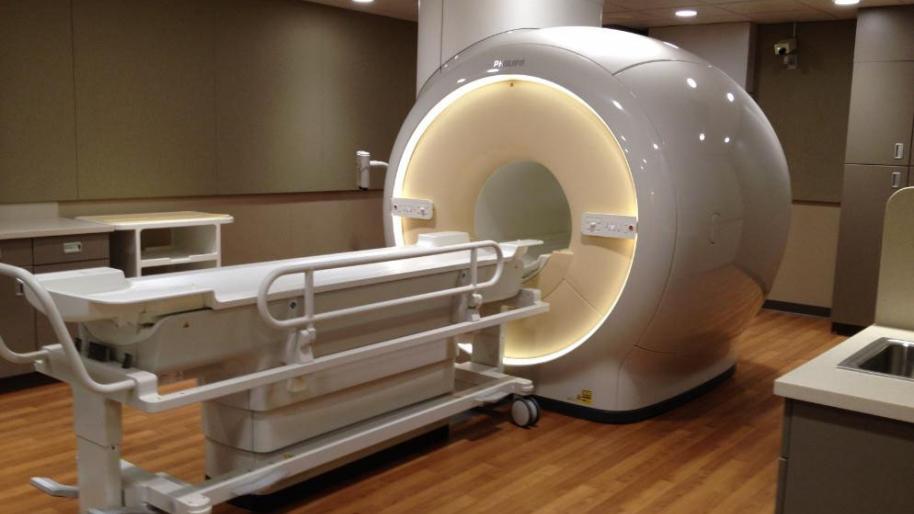
An MRI exam is painless. You don't feel the magnetic field or radio waves. Most MRI machines consist of a large magnet shaped like a tunnel. You lie on a table that slides into the tunnel. A computer creates a composite, three-dimensional representation of your body. Two-dimensional images are then created and displayed on a monitor and/or converted into photographic film for further viewing and analysis. Through clinical research, the Radiology Department has taken a leadership role in establishing new MRI techniques that distinguish benign from malignant tumors in the kidney, liver and adrenal glands as well as MR angiography (to view major blood vessels) and MR cholangiopancreatography (to help diagnose bile duct and pancreatic disorders).