Childhood Astrocytomas Treatment (PDQ®): Treatment - Patient Information [NCI]
General Information About Childhood Astrocytomas
Childhood astrocytomas are tumors that start in the star-shaped brain cells called astrocytes.
An astrocyte is a type of glial cell. Glial cells hold nerve cells in place, bring food and oxygen to them, and help protect them from disease, such as infection. Gliomas are tumors that form from glial cells. An astrocytoma is a type of glioma.
Astrocytoma is the most common type of glioma diagnosed in children. It can form anywhere in the central nervous system (brain and spinal cord).
This summary is about the treatment of tumors that begin in astrocytes in the brain (primary brain tumors). Metastatic brain tumors are formed by cancer cells that begin in other parts of the body and spread to the brain. Treatment of metastatic brain tumors is not discussed here.
Brain tumors can occur in both children and adults. However, treatment for children may be different than treatment for adults. See the following PDQ summaries for more information about other types of brain tumors in children and adults:
- Childhood Brain and Spinal Cord Tumors Treatment Overview
- Adult Central Nervous System Tumors Treatment
Astrocytomas may be low-grade or high-grade.
Low-grade brain tumors grow and press on nearby areas of the brain. They rarely spread into other tissues. High-grade brain tumors are likely to grow quickly and spread into other brain tissue. When a tumor grows into or presses on an area of the brain, it may stop that part of the brain from working the way it should. Both low-grade and high-grade brain tumors can cause signs and symptoms and almost all need treatment.
The central nervous system controls many important body functions.
Astrocytomas are most common in these parts of the central nervous system (CNS):
- Cerebrum: The largest part of the brain, at the top of the head. The cerebrum controls thinking, learning, problem-solving, speech, emotions, reading, writing, and voluntary movement.
- Cerebellum: The lower, back part of the brain (near the middle of the back of the head). The cerebellum controls voluntary movement, balance, and posture.
- Brain stem: The part that connects the brain to the spinal cord, in the lowest part of the brain (just above the back of the neck). The brain stem controls breathing, heart rate, and the nerves and muscles used in seeing, hearing, walking, talking, and eating.
- Hypothalamus: The area in the middle of the base of the brain. It controls body temperature, hunger, and thirst.
- Visual pathway: The group of nerves that connect the eye with the brain.
- Spinal cord: The column of nerve tissue that runs from the brain stem down the center of the back. It is covered by three thin layers of tissue called membranes. The spinal cord and membranes are surrounded by the vertebrae (back bones). Spinal cord nerves carry messages between the brain and the rest of the body, such as a message from the brain to cause muscles to move or a message from the skin to the brain to feel touch.
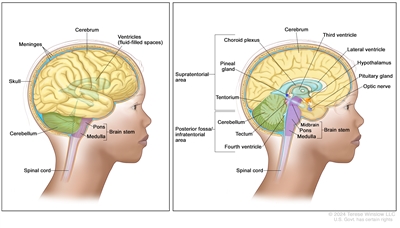
Anatomy of the brain. The supratentorial area (the upper part of the brain) contains the cerebrum, lateral ventricle and third ventricle (with cerebrospinal fluid shown in blue), choroid plexus, pineal gland, hypothalamus, pituitary gland, and optic nerve. The posterior fossa/infratentorial area (the lower back part of the brain) contains the cerebellum, tectum, fourth ventricle, and brain stem (midbrain, pons, and medulla). The tentorium separates the supratentorium from the infratentorium (right panel). The skull and meninges protect the brain and spinal cord (left panel).
The cause of most childhood brain tumors is not known.
Anything that increases your risk of getting a disease is called a risk factor. Having a risk factor does not mean that you will get cancer; not having risk factors doesn't mean that you will not get cancer. Talk with your child's doctor if you think your child may be at risk. Possible risk factors for astrocytoma include:
- Past radiation therapy to the brain.
- Having certain genetic disorders, such as neurofibromatosis type 1 (NF1) or tuberous sclerosis.
The signs and symptoms of astrocytomas are not the same in every child.
Signs and symptoms depend on the following:
- Where the tumor forms in the brain or spinal cord.
- The size of the tumor.
- How fast the tumor grows.
- The child's age and development.
Some tumors do not cause signs or symptoms. Signs and symptoms may be caused by childhood astrocytomas or by other conditions. Check with your child's doctor if your child has any of the following:
- Morning headache or headache that goes away after vomiting.
- Nausea and vomiting.
- Vision, hearing, and speech problems.
- Loss of balance and trouble walking.
- Worsening handwriting or slow speech.
- Weakness or change in feeling on one side of the body.
- Unusual sleepiness.
- More or less energy than usual.
- Change in personality or behavior.
- Seizures.
- Weight loss or weight gain for no known reason.
- Increase in the size of the head (in infants).
Tests that examine the brain and spinal cord are used to detect (find) childhood astrocytomas.
The following tests and procedures may be used:
- Physical exam and health history: An exam of the body to check general signs of health. This includes checking for signs of disease, such as lumps or anything else that seems unusual. A history of the patient's health habits and past illnesses and treatments will also be taken.
- Neurological exam: A series of questions and tests to check the brain, spinal cord, and nerve function. The exam checks a person's mental status, coordination, and ability to walk normally, and how well the muscles, senses, and reflexes work. This may also be called a neuro exam or a neurologic exam.
- Visual field exam: An exam to check a person's field of vision (the total area in which objects can be seen). This test measures both central vision (how much a person can see when looking straight ahead) and peripheral vision (how much a person can see in all other directions while staring straight ahead). The eyes are tested one at a time. The eye not being tested is covered.
- MRI (magnetic resonance imaging) with gadolinium: A procedure that uses a magnet, radio waves, and a computer to make a series of detailed pictures of the brain and spinal cord. A substance called gadolinium is injected into a vein. The gadolinium collects around the cancer cells so they show up brighter in the picture. This procedure is also called nuclear magnetic resonance imaging (NMRI). Sometimes magnetic resonance spectroscopy (MRS) is done during the same MRI scan to look at the chemical makeup of the brain tissue.
Childhood astrocytomas are usually diagnosed and removed in surgery.
If doctors think there may be an astrocytoma, a biopsy may be done to remove a sample of tissue. For tumors in the brain, a part of the skull is removed and a needle is used to remove tissue. Sometimes, the needle is guided by a computer. A pathologist views the tissue under a microscope to look for cancer cells. If cancer cells are found, the doctor may remove as much tumor as safely possible during the same surgery. Because it can be hard to tell the difference between types of brain tumors, you may want to have your child's tissue sample checked by a pathologist who has experience in diagnosing brain tumors.
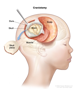
Craniotomy: An opening is made in the skull and a piece of the skull is removed to show part of the brain.
The following test may be done on the tissue that was removed:
- Immunohistochemistry: A laboratory test that uses antibodies to check for certain antigens (markers) in a sample of a patient's tissue. The antibodies are usually linked to an enzyme or a fluorescent dye. After the antibodies bind to a specific antigen in the tissue sample, the enzyme or dye is activated, and the antigen can then be seen under a microscope. This type of test is used to help diagnose cancer and to help tell one type of cancer from another type of cancer. An MIB-1 test is a type of immunohistochemistry that checks tumor tissue for an antigen called MIB-1. This may show how fast a tumor is growing.
- Molecular testing: A laboratory test to check for certain genes, proteins, or other molecules in a sample of blood or bone marrow. Molecular tests also check for certain changes in a gene or chromosome that may cause or affect the chance of developing a brain tumor. A molecular test may be used to help plan treatment, find out how well treatment is working, or make a prognosis.
Sometimes tumors form in a place that makes them hard to remove. If removing the tumor may cause severe physical, emotional, or learning problems, a biopsy is done and more treatment is given after the biopsy.
Children who have a rare genetic condition called NF1 may form a low-grade astrocytoma in the area of the brain that controls vision and may not need a biopsy. If the tumor does not continue to grow or symptoms do not occur, surgery to remove the tumor may not be needed.
Certain factors affect prognosis (chance of recovery) and treatment options.
The prognosis and treatment options depend on the following:
- Whether the tumor is a low-grade or high-grade astrocytoma.
- Where the tumor has formed in the CNS and if it has spread to nearby tissue or to other parts of the body.
- How fast the tumor is growing.
- The child's age.
- Whether cancer cells remain after surgery.
- Whether there are changes in certain genes.
- Whether the child has NF1 or tuberous sclerosis.
- Whether the child has diencephalic syndrome (a condition which slows physical growth).
- Whether the astrocytoma has just been diagnosed or has recurred (come back).
For recurrent astrocytoma, prognosis and treatment depend on how much time passed between the time treatment ended and the time the astrocytoma recurred.
Treatment Option Overview
There are different types of treatment for patients with childhood astrocytoma.
Different types of treatment are available for children with astrocytomas. Some treatments are standard (the currently used treatment), and some are being tested in clinical trials. A treatment clinical trial is a research study meant to help improve current treatments or obtain information on new treatments for patients with cancer. When clinical trials show that a new treatment is better than the standard treatment, the new treatment may become the standard treatment.
Because cancer in children is rare, taking part in a clinical trial should be considered. Some clinical trials are open only to patients who have not started treatment.
Children with astrocytomas should have their treatment planned by a team of health care providers who are experts in treating childhood brain tumors.
Treatment will be overseen by a pediatric oncologist, a doctor who specializes in treating children with cancer. The pediatric oncologist works with other healthcare providers who are experts in treating children with brain tumors and who specialize in certain areas of medicine. These may include the following specialists:
- Pediatrician.
- Pediatric neurosurgeon.
- Neurologist.
- Neuropathologist.
- Neuroradiologist.
- Rehabilitation specialist.
- Radiation oncologist.
- Endocrinologist.
- Psychologist.
Childhood brain tumors may cause signs or symptoms that begin before the cancer is diagnosed and continue for months or years.
Signs or symptoms caused by the tumor may begin before diagnosis. These signs or symptoms may continue for months or years. It is important to talk with your child's doctors about signs or symptoms caused by the tumor that may continue after treatment.
Six types of treatment are used:
Surgery
Surgery is used to diagnose and treat childhood astrocytoma, as discussed in the General Information section of this summary. After surgery an MRI (magnetic resonance imaging) is done to see if any cancer cells remain. If cancer cells are found, further treatment depends on:
- Where the remaining cancer cells are.
- The grade of the tumor.
- The age of the child.
After the doctor removes all the cancer that can be seen at the time of the surgery, some patients may be given chemotherapy or radiation therapy after surgery to kill any cancer cells that are left. Treatment given after the surgery, to lower the risk that the cancer will come back, is called adjuvant therapy.
Observation
Observation is closely monitoring a patient's condition without giving any treatment until signs or symptoms appear or change. Observation may be used:
- If the patient has no symptoms, such as patients with neurofibromatosis type1.
- If the tumor is small and is found when a different health problem is being diagnosed or treated.
- After the tumor is removed by surgery until signs or symptoms appear or change.
Radiation therapy
Radiation therapy is a cancer treatment that uses high-energy x-rays or other types of radiation to kill cancer cells or keep them from growing. External radiation therapy uses a machine outside the body to send radiation toward the area of the body with cancer.
Certain ways of giving radiation therapy can help keep radiation from damaging nearby healthy tissue. These types of radiation therapy include the following:
- Conformal radiation therapy: Conformal radiation therapy is a type of external radiation therapy that uses a computer to make a 3-dimensional (3-D) picture of the tumor and shapes the radiation beams to fit the tumor.
- Intensity-modulated radiation therapy (IMRT): IMRT is a type of 3-dimensional (3-D) external radiation therapy that uses a computer to make pictures of the size and shape of the tumor. Thin beams of radiation of different intensities (strengths) are aimed at the tumor from many angles.
- Stereotactic radiation therapy: Stereotactic radiation therapy is a type of external radiation therapy. A rigid head frame is attached to the skull to keep the head still during the radiation treatment. A machine aims radiation directly at the tumor. The total dose of radiation is divided into several smaller doses given over several days. This procedure is also called stereotactic external-beam radiation therapy and stereotaxic radiation therapy.
- Proton beam radiation therapy: Proton beam radiation therapy is a type of high-energy, external radiation therapy that uses streams of protons (tiny particles with a positive charge) to kill tumor cells. This type of treatment can lower the amount of radiation damage to healthy tissue near a tumor.
The way the radiation therapy is given depends on the type of tumor and where the tumor formed in the brain or spinal cord. External radiation therapy is used to treat childhood astrocytomas.
Radiation therapy to the brain can affect growth and development, especially in young children. For children younger than 3 years, chemotherapy may be given instead, to delay or reduce the need for radiation therapy.
Chemotherapy
Chemotherapy is a cancer treatment that uses drugs to stop the growth of cancer cells, either by killing the cells or by stopping them from dividing. When chemotherapy is taken by mouth or injected into a vein or muscle, the drugs enter the bloodstream and can reach cancer cells throughout the body (systemic chemotherapy). When chemotherapy is placed directly into the cerebrospinal fluid, an organ, or a body cavity such as the abdomen, the drugs mainly affect cancer cells in those areas (regional chemotherapy). Combination chemotherapy is the use of more than one anticancer drug.
The way the chemotherapy is given depends on the type of tumor and where the tumor formed in the brain or spinal cord. Systemic combination chemotherapy is used in the treatment of children with astrocytoma. High-dose chemotherapy may be used in the treatment of children with newly diagnosed high-grade astrocytoma.
High-dose chemotherapy with stem cell transplant
High doses of chemotherapy are given to kill cancer cells. Healthy cells, including blood -forming cells, are also destroyed by the cancer treatment. Stem cell transplant is a treatment to replace the blood-forming cells. Stem cells (immature blood cells) are removed from the blood or bone marrow of the patient or a donor and are frozen and stored. After the patient completes chemotherapy, the stored stem cells are thawed and given back to the patient through an infusion. These reinfused stem cells grow into (and restore) the body's blood cells.
For high-grade astrocytoma that has come back after treatment, high-dose chemotherapy with stem cell transplant is used if there is only a small amount of tumor.
Targeted therapy
Targeted therapy is a type of treatment that uses drugs or other substances to identify and attack specific cancer cells. Targeted therapies usually cause less harm to normal cells than chemotherapy or radiation therapy do.
There are different types of targeted therapy:
- Monoclonal antibodies: Monoclonal antibodies are immune system proteins made in the laboratory to treat many diseases, including cancer. As a cancer treatment, these antibodies can attach to a specific target on cancer cells or other cells that may help cancer cells grow. The antibodies are able to then kill the cancer cells, block their growth, or keep them from spreading. Monoclonal antibodies are given by infusion. They may be used alone or to carry drugs, toxins, or radioactive material directly to cancer cells. Bevacizumab is a monoclonal antibody and vascular endothelial growth factor inhibitor that binds to a protein called vascular endothelial growth factor (VEGF) and may prevent the growth of new blood vessels that tumors need to grow. Bevacizumab is used to treat childhood astrocytoma.monoclonal antibodies: how monoclonal antibodies treat cancerHow do monoclonal antibodies work to treat cancer? This video shows how monoclonal antibodies, such as trastuzumab, pembrolizumab, and rituximab, block molecules cancer cells need to grow, flag cancer cells for destruction by the body's immune system, or deliver harmful substances to cancer cells.
- Protein kinase inhibitors work in different ways. There are several kinds of protein kinase inhibitors.
- mTOR inhibitors: This treatment stops the protein that helps cells divide and survive. Everolimus and sirolimus are mTOR inhibitors used to treat childhood subependymal giant cell astrocytomas.
- BRAF inhibitors: This treatment blocks the activity of proteins needed for cell growth and may kill cancer cells. The BRAF gene is found in a mutated (changed) form in some gliomas and blocking it may help keep cancer cells from growing. The combination of the BRAF inhibitors dabrafenib and trametinib are being studied to treat newly diagnosed low-grade astrocytomas or high-grade astrocytomas that have recurred or stopped responding to treatment. Dabrafenib and trametinib given after radiation are also being studied to treat newly diagnosed high-grade gliomas.
- MEK inhibitors: This treatment blocks proteins needed for cell growth and may kill cancer cells. The MEK inhibitor is being studied to treat low-grade astrocytoma that has recurred or stopped responding to treatment. It is also being studied in combination with chemotherapy to treat newly diagnosed NF1-associated low-grade gliomas.
See Drugs Approved for Brain Tumors for more information.
New types of treatment are being tested in clinical trials.
This summary section describes treatments that are being studied in clinical trials. It may not mention every new treatment being studied. Information about clinical trials is available from the NCI website.
Immunotherapy
Immunotherapy is a treatment that uses the patient's immune system to fight cancer. Substances made by the body or made in a laboratory are used to boost, direct, or restore the body's natural defenses against cancer. This cancer treatment is a type of biologic therapy.
- Immune checkpoint inhibitor therapy: Some types of immune cells, such as T cells, and some cancer cells have certain proteins, called checkpoint proteins, on their surface that keep immune responses in check. When cancer cells have large amounts of these proteins, they will not be attacked and killed by T cells. Immune checkpoint inhibitors block these proteins and the ability of T cells to kill cancer cells is increased.
- PD-1 and PD-L1 inhibitor therapy: PD-1 is a protein on the surface of T cells that helps keep the body's immune responses in check. PD-L1 is a protein found on some types of cancer cells. When PD-1 attaches to PD-L1, it stops the T cell from killing the cancer cell. PD-1 and PD-L1 inhibitors keep PD-1 and PD-L1 proteins from attaching to each other. This allows the T cells to kill cancer cells. PD-1 inhibitors are being studied to treat high-grade astrocytoma that has recurred.
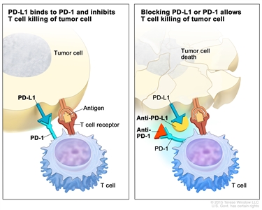
Immune checkpoint inhibitor. Checkpoint proteins, such as PD-L1 on tumor cells and PD-1 on T cells, help keep immune responses in check. The binding of PD-L1 to PD-1 keeps T cells from killing tumor cells in the body (left panel). Blocking the binding of PD-L1 to PD-1 with an immune checkpoint inhibitor (anti-PD-L1 or anti-PD-1) allows the T cells to kill tumor cells (right panel).
If fluid builds up around the brain and spinal cord, a cerebrospinal fluid diversion procedure may be done.
Cerebrospinal fluid diversion is a method used to drain fluid that has built up around the brain and spinal cord. A shunt (long, thin tube) is placed in a ventricle (fluid-filled space) of the brain and threaded under the skin to another part of the body, usually the abdomen. The shunt carries extra fluid away from the brain so it may be absorbed elsewhere in the body.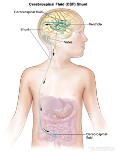
Cerebrospinal fluid (CSF) diversion. Extra CSF is removed from a ventricle in the brain through a shunt (tube) and is emptied into the abdomen. A valve controls the flow of CSF.
Treatment for childhood astrocytomas may cause side effects.
For information about side effects that begin during treatment for cancer, see our Side Effects page.
Side effects from cancer treatment that begin after treatment and continue for months or years are called late effects. Late effects of cancer treatment may include the following:
- Physical problems that affect the following:
- Vision problems, including blindness.
- Blood vessel problems.
- Endocrine function.
- Changes in mood, feelings, thinking, learning, or memory.
- Second cancers (new types of cancer).
Some late effects may be treated or controlled. It is important to talk with your child's doctors about the effects cancer treatment can have on your child. (See the PDQ summary on Late Effects of Treatment for Childhood Cancer for more information.)
Patients may want to think about taking part in a clinical trial.
For some patients, taking part in a clinical trial may be the best treatment choice. Clinical trials are part of the cancer research process. Clinical trials are done to find out if new cancer treatments are safe and effective or better than the standard treatment.
Many of today's standard treatments for cancer are based on earlier clinical trials. Patients who take part in a clinical trial may receive the standard treatment or be among the first to receive a new treatment.
Patients who take part in clinical trials also help improve the way cancer will be treated in the future. Even when clinical trials do not lead to effective new treatments, they often answer important questions and help move research forward.
Patients can enter clinical trials before, during, or after starting their cancer treatment.
Some clinical trials only include patients who have not yet received treatment. Other trials test treatments for patients whose cancer has not gotten better. There are also clinical trials that test new ways to stop cancer from recurring (coming back) or reduce the side effects of cancer treatment.
Clinical trials are taking place in many parts of the country. Information about clinical trials supported by NCI can be found on NCI's clinical trials search webpage. Clinical trials supported by other organizations can be found on the ClinicalTrials.gov website.
Follow-up tests may be needed.
Some of the tests that were done to diagnose the cancer or to find out the stage of the cancer may be repeated. (See the General Information section for a list of tests.) Some tests will be repeated in order to see how well the treatment is working. Decisions about whether to continue, change, or stop treatment may be based on the results of these tests.
Regular MRIs will continue to be done after treatment has ended. The results of the MRI can show if your child's condition has changed or if the astrocytoma has recurred (come back). If the results of the MRI show a mass in the brain, a biopsy may be done to find out if it is made up of dead tumor cells or if new cancer cells are growing.